References

American Society of Anaesthesiologists. ASA Physical Status Classification System. 2014. https://www.asahq.org/resources/clinical-information/asa-physical-status-classification-system (accessed 20th January 2019)

Fraser M, Girling S. First aid and emergency procedures. In: Fraser M, Girling S Chichester: Wiley Blackwell; 2009

Katz J, Clarke H, Seltzer Z. Preventive analgesia: quo vadimus?. Anesth Analg. 2011; 113

Keller LS, Griffin JW, Lang CM. Reproductive failure associated with cystic rete ovarii in Guinea pigs. Veterinary Pathology. 1987; 1:(24)335-9

Lennox AM. Monitoring anaesthetics in exotics. The Veterinary Nurse. 2014; 5:(6)306-11

Mayer J, Donnelly T. Section 1: Diseases and disorders. In: Mayer J, Donnelly T (eds). Missouri: Elsevier; 2013

Orr H. Rodents: Neoplastic and endocrine disease. In: Keeble E, Meredith A (eds). Gloucester: British Small Animal Veterinary Association; 2011

Cystic ovarian disease. 2018. https://www.vetstream.com/treat/exotis/guinea-pigs/diseases/cystic-ovarian-disease (accessed 17th January, 2019)

Pilny A. Ovarian cystic disease in guinea pigs. Vet Clin North Am Exot Anim Pract. 2014; 17:(1)69-75 https://doi.org/10.1016/j.cvex.2013.09.003

Guinea pig reproduction basics. 2017. https://lafeber.com/vet/guinea-pig-reproduction-basics/ (accessed 31st December, 2018)

Schuetzenhofer G, Goericke-Pesch S, Wehrend A. Effects of deslorelin implants on ovarian cysts in guinea pigs. Schweiz Arch Tierheilk. 2011; 153:416-17 https://sat.gstsvs.ch/fileadmin/media/pdf/archive/2011/09/SAT153090416.pdf

Woolf CJ, Wall PD. Morphine-sensitive and morphine-insensitive actions of C-fibre input on the rat spinal cord. Neurosci Lett. 1986; 64:221-5

Cystic ovarian disease in female guinea pigs
02 July 2019
9 mins read
Volume 10 · Issue 6

Abstract

This article will look at cystic ovaries in female guinea pigs. Cystic ovaries can be functional or non-functional fluid filled cysts that usually develop spontaneously in the older sow. The presence of cysts usually reduces fertility and potentially causes serious uterine disease. Identifying common symptoms related to this condition can aid the veterinary nurse when performing clinical examinations. Species specific care is vital to securing optimum patient care and the chance of a good outcome.

Veterinary nurses (VNs) working in referral or first opinion practice carry out routine physical examinations of patients daily. This can be as simple as taking a temperature, checking mucous membrane colour or performing a full health check as part of a nursing consultation. As prey species, guinea pigs instinctively hide signs of pain/disease and injury, therefore at every opportunity a clinical examination is prudent. This, together with a sound knowledge of common diseases and conditions in this species will provide the cornerstone to gold standard nursing care and improvement of clinical satisfaction and client trust.

Reproductive pathophysiology

Cystic ovarian disease is very common in sows over the age of 1.5 years, an incidence of 76% has been reported at post mortem (Keller et al, 1987). Puberty occurs at around 2 months of age in females and peak reproduction is between 3 and 20 months, although some sows may continue to breed until 4–5 years of age. Guinea pigs are seasonally polyoestrous but breed year round in captivity. The sow possesses a single pair of inguinal mammary glands, paired ovaries, a bicornuate uterus consisting of paired uterine horns, a uterine body and a single cervix (Pollock, 2017).

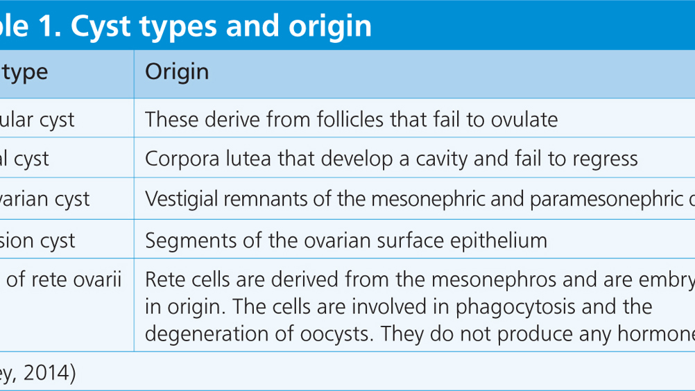
Five types of cysts have been identified in the ovaries of various laboratory animals and are classified according to their tissue of origin within the ovary (Table 1). The three most common cysts in guinea pigs in order of prevalence are: cystic rete ovarii; follicular cysts; and parovarian cysts (Pilny, 2014).


Table 1. Cyst types and origin
Cyst type Origin
Follicular cyst These derive from follicles that fail to ovulate
Luteal cyst Corpora lutea that develop a cavity and fail to regress
Parovarian cyst Vestigial remnants of the mesonephric and paramesonephric ducts
Inclusion cyst Segments of the ovarian surface epithelium
Cysts of rete ovarii Rete cells are derived from the mesonephros and are embryonic in origin. The cells are involved in phagocytosis and the degeneration of oocysts. They do not produce any hormones
(Pilney, 2014)

Physical examination and symptoms

Prey species are masters of disguising pain and illness, this often means symptoms are mild or undetectable by owners even when significant disease processes are present. Unlike rabbits, guinea pigs do not require annual vaccination in the UK so there is no compelling reason for clients to bring guinea pigs to the practice. One of the main reasons for a ‘routine visit’ to the practice for a guinea pig is nail clipping. It is imperative that VNs and veterinary surgeons (VS) use this opportunity to examine the patient and encourage the owner to discuss any concerns.

Usually, the most common clinical sign observed by owners in regards to cystic ovarian disease is non-pruritic, bilateral, symmetrical alopecia, which tends to be found over both flanks (Figure 1). The skin is usually normal in appearance in these areas, however there may be epitheliomatous degeneration around the nipples (Figure 2). External genitalia may be enlarged, and masses of varying size may be palpated in the abdomen (Orr, 2011). Typical to their species, guinea pigs may also present with very non-specific signs of illness including: loss of body condition; appetite loss; lethargy; abdominal pain or vocalisation when being handled. Some guinea pigs may be completely asymptomatic and smaller cysts may be found incidentally during routine neutering for example.

Figure 1. Middle aged sow demonstrating non-pruritic alopecia over flanks.
Figure 2. Epitheliomatous degeneration around nipples of sow.

Diagnosis

To aid diagnosis a VS may use ultrasonography to detect ovarian cysts. This usually reveals a fluid filled component of one or both the ovaries and is useful to distinguish between cyst or tumour (Pollock, 2017). Radiography can also be used but diagnosis may be more challenging as cystic tissue has poor radiographic opacity making differentiating between other organs difficult, especially if they are less than 2 cm in diameter (Perpinan and Johnson-De-laney, 2018). If ultrasonography is not available, a presumptive diagnosis may be made by a VS based on clinical signs and examination and diagnosis achieved by performing an exploratory laparotomy. Informed consent must be gained from the owner, and a risk versus benefit scenario discussed with the veterinary team.

Surgical treatment

Current thought concludes that ovariectomy or ovariohysterectomy (Figure 3) is the treatment of choice for this condition (Orr, 2011). If surgery is successful it is curative, what needs to be considered is the candidate for surgery. The candidate in question is usually an older prey animal with at least one known significant disease process. Many of the guinea pigs presenting for surgery due to cystic ovarian disease can be scored at least 3 on the physical status scale designed by the American Society of Anesthesiologists (Table 2) — meaning they are at the very least, a moderate anaesthetic risk. Many will score higher and pose a much higher threat, especially if there is concurrent disease.

Figure 3. Demonstration of bilateral ovarian cysts post ovariohysterectomy in 4-year-old sow.

Table 2. Anaesthetic risk groups
Risk level 1 — minimal risk Normal, healthy animal with no underlying disease
Risk level 2 — slight risk Mild systemic disturbance but able to compensate
Risk level 3 — moderate risk Moderate systemic disease with clinical signs and/or observable pathology
Risk level 4 — high risk Significantly compromised by one or multiple disease processes
Risk level 5 — severe risk Animal is moribund and not expected to survive without immediate intervention
Adapted from the American Society of Anesthesiologists, 2014

An alternative to ovariohysterectomy is an ovariectomy, this is usually reserved for cases where there is no evidence of concurrent uterine disease. This can be performed via incisions on the dorsal flanks of the guinea pig, ventral to the erector spinae muscle. Blunt dissection through the muscle will provide access to the ovary, which can be slightly exteriorised and aseptic drainage performed with direct visualisation. This can be helpful if the cyst is too large to be extracted from the incision. In dogs, ovariectomy versus ovariohysterectomy results in lower complication rates, and in guinea pigs, advantages of ovariectomy include small and dorsal incisions, less gastrointestinal disruption/handling and quicker surgical time (Pilny, 2014).

VNs are often called on to monitor anaesthesia in a huge range of species, sometimes without prior experience and limited knowledge. Unfamiliarity with normal/abnormal vital signs, monitoring techniques and inability to achieve intubation and vascular access are all contributing factors to patient demise (Lennox, 2014).

Surgical/anaesthetic nursing considerations

There are a number of considerations for nursing the guinea pig during surgery and anaesthesia:

  • Ensure the patient is stable before surgery. This is of the greatest importance in exotic species. Admitting the animal prior to surgery to commence supportive feeding, administration of pain relief, supportive medication and fluid therapy to correct hydration deficits are vital to increase the chance of a favourable outcome.
  • VNs and VSs should become familiar with guinea pig vital signs, anatomy, anaesthetic protocols and monitoring methods before beginning surgery.
  • Provide pre-emptive analgesia under veterinary direction — prey animals hide the extent of their pain pre and post-operatively. As in other species, experiments demonstrate that administration of an analgesic prior to an acute pain stimulus will more effectively minimise changes associated with central sensitisation than when the same analgesic is given after pain has been established (Woolf and Wall, 1986). Also, although patients may be unconscious through general anaesthesia, the processes leading to sensitisation of spinal and medullary dorsal horn neurons remain intact (Katz et al, 2011); this means the physiological response to pain can still be observed even under general anaesthesia, and will most certainly become apparent to the patient as soon as consciousness is regained.
  • Patient positioning — herbivorous small mammals have a large abdominal:thoracic ratio and large ovarian cysts increase pressure on the diaphragm which can result in respiratory compromise. As small herbivores such as guinea pigs and rabbits rely on flattening of the diaphragm rather than outward movement of the ribcage, lifting the thorax during surgery should aid the patient's respiration (Fraser and Girling, 2009).
  • Intubation — this is very difficult to achieve in guinea pigs. Guinea pigs possess a palatal ostium which is a hole in the soft palate that connects the oropharynx with the rest of the pharynx. This can be traumatised easily when intubation is attempted, therefore a facemask is the most common method used to deliver oxygen and anaesthetic agent in general practice. The difficulty in intubating quickly and safely likely contributes towards the higher anaesthetic mortality rates seen in this species.
  • Hypothermia — small mammals have a huge surface area to bodyweight ratio. In practical terms this means they can become hypo and hyperthermic more readily than cats and dogs. From the moment pre-medication or anaesthesia is induced, metabolic rates and body temperature will fall. Normally when an animal is hypothermic the body initiates vasoconstriction of the peripheral vessels, however this reflex is neutralised if vasodilating agents such as isoflurane or sevoflurane are used. As core temperature falls, the sino-atrial node beats more slowly, there is a drop in cardiac output and blood pressure, and emergency drugs such as atropine and glycopyrrolate are therefore unlikely to effectively correct bradycardia. As temperatures approach 32°C, asystole or fibrillation may occur spontaneously (Robertson, 2015). Metabolism is slowed and liver function is impaired, delaying breakdown of anaesthetic drugs, which prolongs recovery times. In human studies, intra-operative hypothermia has been linked to increased incidence of postoperative wound infection. Results concluded this was the result of poor perfusion to the periphery, vasoconstriction and low oxygen concentration at the surgical site (Robertson, 2015). To aid with patient thermoregulation, forced air warming devices, hot hands, heat pads, incubators, foil and bubble wrap can all be utilised.
  • Fluid therapy is imprecise, mostly because the volumes of various body compartments are constantly changing. Therefore, only an estimate of the volumes required are administered and in small exotic mammals, which have a high basal requirement, a three stage technique should be employed as deemed necessary by a VS.
  • Resuscitation — the aim of the resuscitation period of fluid therapy is to restore tissue perfusion and oxygenation. Resuscitation fluids require to be utilised in cases of hypovolaemia which is a reasonable concern in these surgical patients. Relatively large volumes of tissue removal, evaporation from the abdomen and a higher risk of haemorrhage in diseased tissue are contributing factors. In the author's experience it is impractical to use subcutaneous fluids at resuscitation level and this highlights the need for vascular access. An intravenous catheter can usually be placed in the cephalic or lateral saphenous of adult guinea pigs (Figure 4). An alternative would be the introduction of intraperitoneal or intraosseous fluids, but there are limitations to both, and discussion is outside the scope of this article.
  • Hydration — this is restoration of interstitial fluid
  • Maintenance — this is where ongoing losses are maintained and managed.
Figure 4. Access to the left cephalic vein in an adult guinea pig.

Postoperative care should include recovery in a warm, quiet environment away from predators, the provision of good analgesia, supportive feeding as required and the administration of fluids and supportive medications. Keeping patient hospital sheets up to date is extremely important and comprehensive nursing handovers ensure patient care continuity in a species where accurate dosing is of the utmost importance.

Medical management

In scenarios where the patient is unable to undergo surgery, medical management can be attempted by the VS. It should however, be made clear to the owner the results will be temporary and patient re-evaluation ongoing. Forms of medical management include:

  • Human chorionic gonadotrophin (HCG) — 1000iu/guinea pig intramuscularly. At this dosage the volume will be approximately 1 ml which is a large volume to inject intramuscularly in a guinea pig. It is also important to note HCG will stimulate an antibody response, potentially making subsequent doses less effective and possibly stimulating an allergic reaction with repeated injections (Mayer and Donnelly, 2013).
  • Gonadotropin-releasing hormone (GnRH) — this drug is used to treat ovarian cysts in cattle and induce oestrous in cats. Unlike HCG it does not stimulate an immune response — the recommended dose is 25 ug/guinea pig every 2 weeks for two injections.
  • Deslorelin implant (an injectable GnRH superagonist) — according to Schuetzenhofer et al (2011) treatment with slow release GnRH did not influence the size of ovarian cysts in coincidentally selected guinea pigs and it is thought the localisation and physiological activity of GnRH and its receptors in guinea pigs differ from other mammalian GnRH.

Unfortunately cysts of rete ovarii and parovarian cysts are unlikely to respond to hormonal therapy. Therefore percutaneous drainage is the only real alternative therapy to attempt to relieve clinical signs, albeit temporarily. Follicular cysts may respond to medical management and intermittent drainage.

Follow-up consultations can be performed by VNs under veterinary direction in medical cases to check response to treatment and allow the client further time to discuss the patient. The consultation may include a weight check, assessment of the patient's body condition, demeanour, nutritional needs, assessment of pain (observation or abdominal palpation) and discussion with the client regarding the patient's response. The nursing consultation can be used to ascertain ongoing treatment response including improvement of clinical signs or the need to refer back to a VS.

Conclusion

To aid the VS, owner and patient, VNs can provide proactive and forward-thinking nursing care to guinea pigs. Becoming familiar with subtle signs of pain in prey species, providing a low stress environment, careful handling, pre-emptive supportive treatment and a keen eye for early signs of deterioration could make the difference between life and death. Accurate history taking, a thorough clinical examination to identify any abnormalities and swift referral to a VS will not only increase the chances of a better prognosis for the patient, but provide clients with comfort and trust their pet is in the hands of a competent VN. A positive, can do attitude to small mammal nursing will not only increase knowledge and experience, but may break the veterinary taboo of what is expected and achieved in the nursing of exotic patients.

KEY POINTS

  • Cystic ovaries are common in sows over 1.5 years of age and can grow to appreciable sizes.
  • Cysts of rete ovarii are the most common cysts found in female guinea pigs.
  • Diagnosis can be achieved through a combination of abdominal palpation, ultrasonography, radiography or exploratory laparotomy.
  • Cysts can be treated surgically via ovariohysterectomy or ovariectomy or medically using injectable hormones and/or percutaneous drainage.
  • Guinea pigs require species specific nursing care and close anaesthetic monitoring should they become surgical candidates.