Awound is an interruption of anatomic, physiologic, and functional integrity of the body's tissue (Hosgood, 2003). The healing process begins immediately after injury by means of a complex and finely orchestrated continuum of stages (i.e. inflammatory, debridement, repair, and maturation) (Swaim and Henderson, 1997). The process involves sophisticated synchronization of molecular and biochemical events at the cellular level, resulting in a healed wound (Gregory, 1999). Malfunction of any component of the process, or interruption of any stage, may result in delayed healing and chronic or non-healing wounds.
Wounds that do not heal ‘normally’ can be very dif-ficult and stressful to treat. In treating wounds there is no one ‘technique’ or ‘method’ that can be applied to every single wound. Complications of wound healing may arise through:
Knottenbelt (2003) listed 12 factors which may complicate or delay the wound healing in open wounds (Box 1). Infected wounds will heal slower than non-infected wounds and the presence of bacterial organisms within a wound delays healing. Movement will disrupt newly formed capillary buds and increase collagen deposition resulting in a ‘chronic’ state of healing. Foreign material, which can include sand or grit particles, wood, plant matter, metal, glass, will delay healing, as will necrotic tissue, including skin, tendon, bone, muscle etc. For this reason it is useful to allow natural demarcation (an obvious border between healthy and non-viable tissue) to become evident before wound closure is performed. Altered local pH will also affect healing; certain bacteria will result in an acidic or alkaline wound environment, whereas the ideal pH of the wound environment for optimal healing is near neutral. Poor blood supply also has an affect and can occur as a result of major vessel disruption, thrombosis, oedema or contusion, damage to microcirculation, anaemia or delay in capillary formation. Poor oxygen supply may occur for a number of reasons, such as lowered circulating oxygen due to reduced blood flow, and will have an effect on wound healing. Poor nutritional and health status are also important factors; a lack of nutrition will severely reduce the rate of wound healing. Local factors, such as in wounds which are poorly drained, e.g. excessive dead space in a surgically closed wound, may well fail to heal, or will heal far slower than expected. Genetic factors also play a role; in equine wounds certain genetic lines and certain individuals heal less well than others, this is not commonly seen in small animal wounds. Some horses may also have congeni-tally weakened skin which is particularly fragile and likely to be damaged more easily than normal. Cell transformation is also important; certain horses can develop sarcoid transformation at wound sites. These sarcoids require removal in order for wound healing to continue.
Other factors which the author considers when faced with a delay in wound healing include: biofilms; tension; bandaging/casts; seroma/hae-matoma formation; and concurrent medication.
When dealing with a non-healing wound veterinary nurses should consider whether any of these factors may be the cause of the delay in the normal wound healing process.
Infection
Infection delays wound healing. If infection is thought to be the cause of the delay in wound healing in a surgical wound, then a thorough review of the patient, the environment and surgical technique should be carried out.
Bacterial, granulocyte, and macrophage colla-genases degrade collagen, thereby decreasing wound strength (Hosgood, 2003). In addition, decreased fi-broblast activity during infection has a negative effect on healing (Hosgood, 2003). The end result is a tendency for wound disruption. It is vitally important to use thorough initial wound management techniques, such as lavage and debridement, along with systemic and topical medication, in order to remove non-viable tissue and infection and prevent wound disruption.
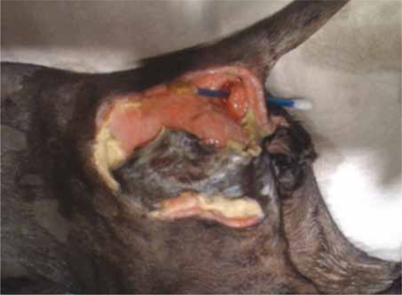
A pathogenic organism, such as meticillin resistant Staphylococcus aureus (MRSA), meticillin resistant Staphylococcus pseudintermedius (MRSP), Pseu-domonas aeruginosa or Proteus spp, may be involved, so ideally a deep tissue culture or biopsy should be taken for aerobic and anaerobic culture and sensitivity testing (Friend, 2009) (Figure 1). If surface samples are collected via a swab it is likely that only the bacteria present in the ‘bioflm’ on the surface of the wound will be collected. In relation to non-healing and infected wounds, the concept of biofilms has a very sig-nificant role to play (Percival and Rogers, 2005) and research is on-going in this area as biofilms are being demonstrated to act as a barrier to antimicrobial penetration (Stewart, 1996; Cochran et al, 2000).
Movement and pressure
Wounds over joints present a challenge to healing in that they are subject to tension, compression, or shearing forces. The desired result of wound healing is for the two sides of a wound to heal together. If they are exposed to these forces, however, healing is impaired. Wounds over extension surfaces of joints (e.g. carpus, stifle) are subject to tension when joint flexion pulls wound edges apart. Thus, meticulous closure is necessary. Casting or splinting the joint is necessary to prevent joint flexion for proper healing (Campbell, 2006).
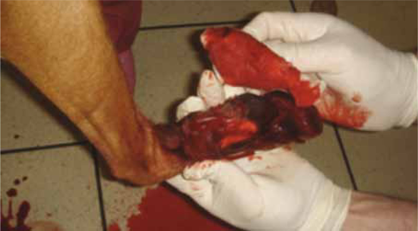
Foot pads are shock absorbing and spread as weight is applied. Thus, in the presence of an open wound, edges are pushed apart, impeding healing (Swaim et al, 1992; Swaim et al, 2003). If sutures are present in the pad, such pad spreading results in sutures tearing through the tissues (Swaim et al, 2003). It is therefore necessary to relieve pressure on foot pads to attain adequate healing, especially in large dogs (Figure 2). Various bandaging and splinting techniques have been evaluated as to their efficacy in reducing pressure on digital and metacarpal or metatarsal pads using various forms of foam rubber pads, metal splints, and combinations of these (Swaim et al, 2003).
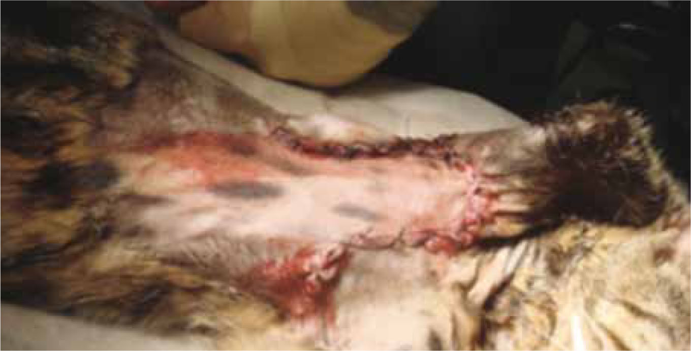
Wounds in the axillary and inguinal areas may result from forelimb entrapment in a collar, vehicular trauma, burns, neoplasia, and infections (Hunt, 1995; Brockman et al, 1996). A primary factor in the impaired healing of such wounds is the shearing movement between the two wound surfaces as the animal ambulates (Swaim and Henderson, 1997). If such wounds have been present for a long period, it is possible that there may be infection with an atypical organism. Thus, a biopsy for culture and sensitivity testing is indicated. Techniques for closing such wounds have included meticulous closure and the use of skin fold flaps, omental pedicle flaps, axial pattern skin flaps (Figure 3), or combinations of these (Hedlund, 2006).
Foreign material and necrotic tissue
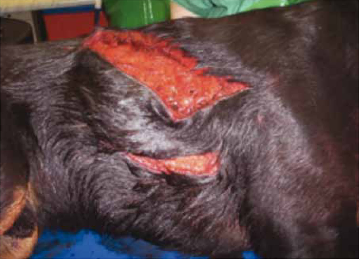
Wounds are much less likely to heal if there is a focus of infection present, this can include material such as debris and dirt within the wound following a road traffic accident, or bone sequestrum following a fracture (Friend, 2009). Such material give bacteria a surface onto which they can adhere, in areas where there is a poor blood supply and low oxygen tension. Due to the poor blood supply, phagocytes are less likely to be able to access these bacteria and additionally systemic antibiotics will have less efficacy due to an inability to penetrate the tissues. These factors highlight the importance of adequate lavage, exploration and debridement of all wounds. All obviously necrotic tissue should be removed from the wound bed via debridement and this may be done as a ‘staged’ process, particularly if the wound is located close to structures such as vessels, nerves etc, which need to be preserved. Wound debridement can be carried out by a wide number of techniques and is a step which should be repeated until the clinician is happy that all foreign material and necrotic tissue have been removed from the wound bed (Figure 4).
Nutrition
Adequate nutritional intake is vital for wound healing to occur, and should be a priority in all critical/ trauma patients. A catabolic state, attributable to malnutrition, is a major contributing factor to non-healing wounds (Amalsadvala and Swaim, 2006). In this condition, the body does not have the necessary protein and energy sources (fats and carbohydrates), therefore, existing stores of protein are broken down to maintain basal functions. This means, the increased calorific and protein demands for healing are not available, and the wound becomes quiescent (Crane, 1989). Glucose and protein are important for normal progression of wound healing. Glucose is the primary source of energy for leukocytes and fibroblasts. It is the integral molecule within the ground substance that is laid down by the fibroblasts. Deposition of this is necessary before collagen formation (Swaim and Henderson, 1997). Thus, glucose deficiency can affect collagen formation and wound strength. Depletion of protein stores can result in attenuated fibroplasia and prolonged healing time (Swaim, 1980). It is vital that patients receive adequate protein levels as they are necessary for animals undergoing healing. Sufficient protein levels help to prevent oedema and promote increased fibroplasia with increased wound strength (Noffsinger et al, 1957).
Vitamins may also affect wound healing. Excess vitamin A labilizes lysosomes to enhance inflamma-tion. Because steroids stabilize lysosomes and inhibit wound repair, vitamin A can counteract this negative effect (Hosgood 2003; Swaim, 1980). Likewise, vitamin E stabilizes lysosomes similar to steroids and thus can inhibit healing in large doses (Hosgood 2003; Swaim, 1980). Again, vitamin A can reverse the effects of vitamin E. Vitamin C deficiency can impair healing in that it is necessary for the hydroxylation of proline and lysine in collagen synthesis (Swaim, 1980; Hosgood 2003). Although dogs and cats do not require exogenous sources of vitamin C, there is the possibility that the vital levels of ascorbic acid in the blood may decrease after trauma (i.e. wounding). Once all other factors affecting wound healing have been ruled out there could be an indication for vitamin C supplementation in these animals (Swaim, 2003).
Zinc deficiency can result in lack of replication of epithelial cells and fibroblasts, causing a weak wound and lack of epithelialization. At the other extreme, an elevated zinc concentration can inhibit macrophag-es, decrease phagocytosis, and interfere with collagen cross linking to have a negative effect on healing (Hosgood 2003; Swaim, 1980), and should be considered once other factors have been excluded.
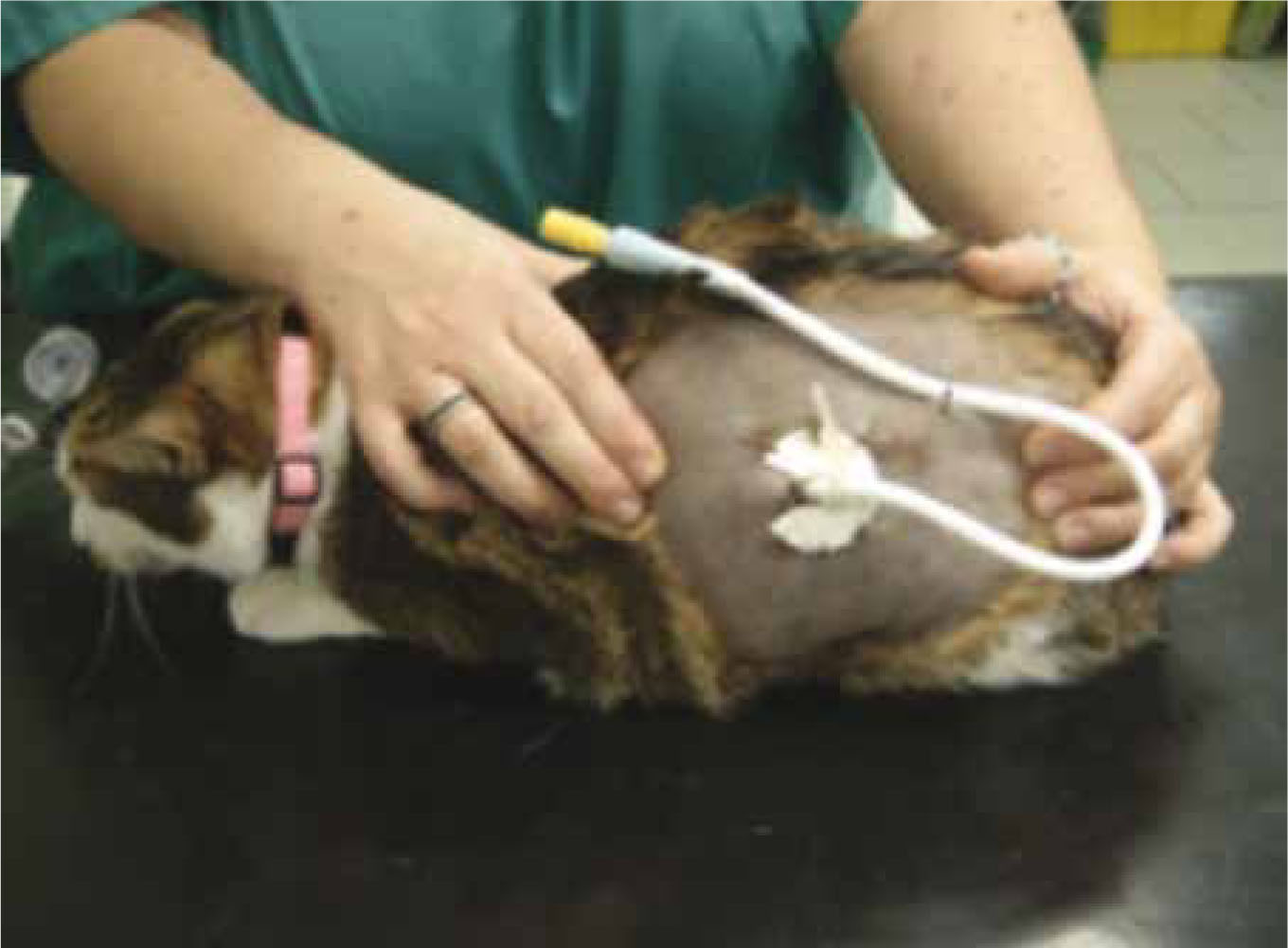
Ideally the patient's nutritional status on admission to the practice should be noted, including a Body Condition Score (BCS) and weight and this should be performed daily during the hospitaliza-tion period. If there is concern that the patient is unable to achieve its resting energy requirement (RER) alone, then assisted feeding techniques such as the placement of oesophogostomy or gastrotomy tubes should be commenced (Figure 5). Many of these patients will require general anaesthesia as part of a wound management protocol, e.g. wound debridement, and so the opportunity for the placement of a feeding tube is likely to be available. For patients requiring sedation or anaesthesia over a prolonged period of time, nutritional status may also need to be addressed in terms of prolonged periods of starvation prior to anaesthesia, and in these cases the patient's calorific requirements should be calculated and compared against the actual calorie intake of the patient.
Additional factors affecting wound healing
There are many other additional factors that may delay wound healing which should also be considered, including medications, e.g. corticosteroids, chemo-therapeutic agents, and underlying disease or conditions (see Table 1). Underlying systemic disease, such as heart disease, may decrease pH and oxygen tension, and interrupt blood supply (Mason, 1993). Mechanical interference by exudate is also a factor that should be considered.
| Diabeteszmellitus |
| Hyperadrenocorticism |
| Infectiouszagents,ze.g.FeLV/FIV |
| Hypothyroidism |
| Anaemia |
| Renalzdisease |
| Liverzdisease |
| Hypoproteinaemia |
| Coagulopathies |
| Neoplasia,e.g.zmast cell tumours |
| Inadequate nutritional status |
Conclusion
Continual reassessment of the wound and its environment is required throughout the wound management process. Changes may be required to the wound management protocol, including reviewing the frequency of dressing changes, changing local management of the wound, altering the patient's environment, e.g. cage rest, to encourage wound healing. In situations where more than one clinician is likely to review the wound, a detailed recording system should be in place so that an accurate re-assessment of the wound can be made, reviewing the condition of the wound, including size, exudate level and appearance, general health of the patient, pain score on removal of dressing etc. Wound management is a role in which nurses can make a true difference; veterinary nurses are often in a position to ensure that wounds heal at their optimum through attention given to both local and systemic factors and they can have a positive influence on the outcome of a case. If veterinary nurses have a good understanding of the wound healing process, this will enable them to correctly assess the stage of wound healing and select appropriate dressing types to optimize the wound environment. A good understanding of factors which may negatively affect wound healing will mean nurses are able to identify these factors as a potential reason for delayed wound healing and make the appropriate changes to the wound management plan.