Hip dysplasia (HD) is a polygenically inherited developmental abnormality that is associated with hip instability and osteoarthritis (OA) (LaFond et al, 2002;Janutta et al, 2006). This affects bone growth and remodelling, resulting in abnormal friction between both joint surfaces and, subsequently, joint deformity and degenerative joint disease (DJD) (Lohi and Nicholas, 2009). Radiographically, a dysplastic animal has a shallow acetabulum and the femoral head fits poorly into it (Kealy and McAllister, 2000).
Breeds likely to develop HD
Both the prevalence and clinical significance of HD vary widely among breeds (Wang et al, 1999;Todhunter et al, 2003), mainly affecting giant and large breed dogs, such as German Shepherds, Labrador Retrievers and Rottweilers (Kealy and McAllister, 2000;LaFond et al, 2002). Greyhounds are rarely affected suggesting that selection in these breeds, which has resulted in good musculoskeletal development, has favourably altered their susceptibility to HD (Mäki et al, 2000).
Factors involved in the development of HD
Several factors besides inheritance have been suggested to play a role in HD development (Silvestre et al, 2007):
Clinical signs
The clinical presentation of dogs with HD (Table 1) is variable and does not correlate with the radiographic changes in joint morphology (Ginja et al, 2008). On the one hand, affected animals can go unnoticed or show clinical signs only at an advanced age (Kapatkin et al, 2002a), on the other hand HD can severely impair locomotive function (Arnbjerg, 1999).
| Lameness |
| Degenerative joint disease |
| Hip laxity and instability |
| Osteoarthritis |
| Synovitis |
| Acetabular microfractures |
| ‘Bunny hopping’ |
| Pain in full extension of the joint |
Hip laxity is the earliest sign of the disease (Kealy and McAllister, 2000), however, the most obvious phenotypic manifestation is DJD of the hip joints (Kealy et al, 2000).
In general, there are two ages at which animals present with overt clinical signs of HD: dogs younger than 1 year of age with hip instability and some articular areas overloading, where pain is caused mainly by tearing or stretching of the round ligament, synovitis and acetabular microfractures; and adult dogs with chronic pain from OA (Manley et al, 2007). Gait abnormalities, such as stiffness, reduced step height, shortened stride length, ‘bunny hopping’, difficulty in rising, climbing stairs or jumping over obstacles are all typical clinical signs. Animals with HD can also show hindlimb lameness and pain in full extension of the joint (Ginja et al, 2008) (Table 1).
Diagnosis of HD
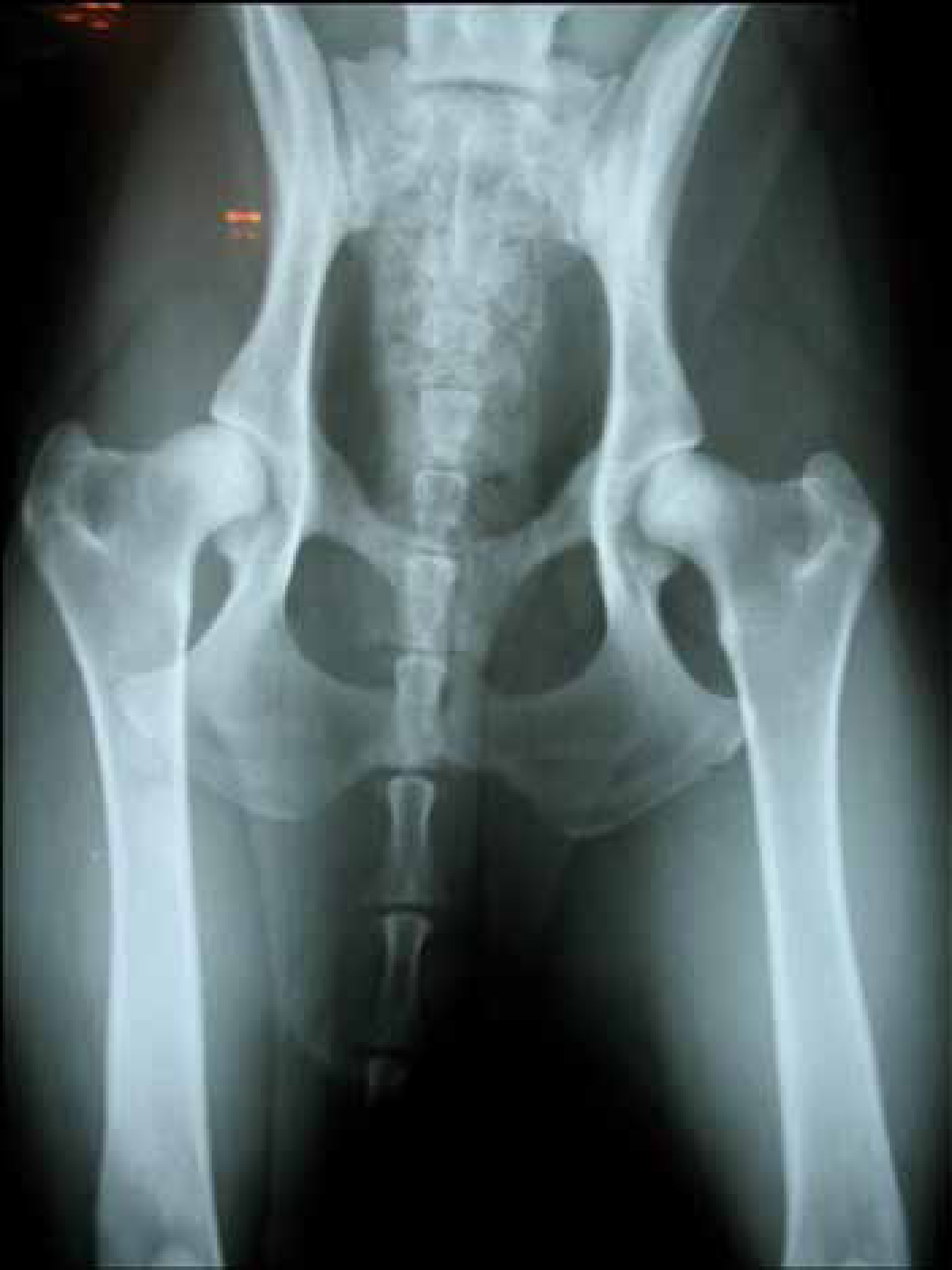
The standard diagnostic method for HD is based on a ventrodorsal hip-extended radiographic view (Figure 1 and Figure 2) and the degree of the disease is classified according to the conventional classification protocols, for example, of the Orthopaedic Foundation for Animals and the British Veterinary Association which are based on joint laxity, subluxation and OA (Mäki et al, 2000). Radiographs are usually helpful in the diagnosis of severe hip OA but are not always beneficial in the diagnosis of mild or moderate disease. In addition, there is no direct correlation between degree of pain and the severity of radiographic changes within the joint (Farrell et al, 2007).

Treatment of dogs with HD
HD can be treated using surgical and conservative methods, the decision on which to use depending on the animal's age, weight, clinical symptoms and the financial situation of the patient's owner (Plante et al, 1997).
Surgery is indicated in older patients, when conservative treatment is not effective, or in young patients, where athletic performance is desired or the owner wishes to slow progression of DJD (Fossum et al, 2002). There are several surgical procedures available:
Conservative management includes medical approach, physiotherapy, dietary changes, weight reduction in obese animals, acupuncture and close confining. The conservative management may be effective in palliating the discomfort associated with HD but will not prevent development and progression of degenerative changes (Manley et al, 2007).
Conservative versus surgical treatment
Although less expensive, conservative treatment does not provide the definitive cure for HD and requires a long period of treatment before it achieves the desired objectives, which are not always possible to reach. It requires owner cooperation and motivation (Bockstahler et al, 2004) for rehabilitation as certain exercises can be performed at home supported by the owners. For this reason, this type of treatment is not advisable for animals whose owners are unable to participate in recovery exercises.
Massage has been shown to reduce pain, increase pain tolerance, and stimulate a release of endorphins and regular massage sessions should be administered by qualified and trained veterinary nurses, under the direction of the veterinary surgeon (Edge-Hughes, 2007).
Thermal agents such as heat or cold are both reported to have pain-relieving effects (Edge-Hughes, 2007). As a general rule, cold therapy (cryotherapy) is indicated during acute inflammation when the lesion exhibits signs of redness, swelling, pain and/or heat. Heat is indicated during the sub-acute and chronic inflammatory phases. These techniques can be used interchangeably (contrast baths), to production of alternating vasoconstriction and vasodilatation of local blood vessels, that stimulate the blood flow (Steiss, 2010).
Conservative treatment is less invasive than surgery and it is known that approximately 75% of young patients treated conservatively reach acceptable clinical function by the time of maturity (Fossum et al, 2002).
Surgical treatment is indicated in elderly patients, in situations where conservative treatment has not been effective, and in young patients when the athletic performance is desired or the owner wants to slow down the DJD progression (Fossum et al, 2002). Surgical treatment is more expensive than conservative treatment; the chosen surgical procedure is dependent on the severity of the joint lesions (Padilha Filho, 1992) and may result in some post-surgical morbidity (Dueland et al, 2001).
Patient management
Management of the HD patient
The veterinary nurse should advise owners to use low fat and protein diets, associated with weight control programmes, and moderate exercise, to prevent obesity and hence the increase of degenerative lesions (Fossum et al, 2002).
A well padded and warm bed in a welcoming environment helps to relieve some of the arthritic pain; non steroidal anti-inflammatory drugs (NSAIDs) can also be administered (Fossum et al, 2002).
It is also important to develop an exercise plan, according to the severity of the symptoms. In general, for animals showing degenerative changes, exercise must be moderate to benefit cartilage nutrition and maintain the muscle mass, which is an important factor for hip stabilization. For example, hydrotherapy associated with other complementary exercises (underwater treadmill) is a good programme for dysplastic animals, as it causes minimal articular stress (Kapatkin et al, 2002b). Aquatic therapy, with temperatures higher than 27°C benefits dogs with arthritic conditions (Saunders, 2007a). Aquatic exercise decreases the weight placed on limbs, therefore increasing the resistance to motion, and providing superficial heat to affected joints. Aquatic therapy has been shown to be effective for human patients with OA by increasing their aerobic capacity and activity levels (Bunning et al, 1991).
Heat therapy increases blood flow, enzymatic activity, and collagen extensibility and leads to muscle relaxation and temporary pain relief in arthritic patients; this therapy can be used to decrease joint inflexibility (Oosterveld et al, 1994).
Gross muscle strengthening can include leash-walking in a tight ‘figure 8’ pattern, underwater treadmill walking, sit to stand exercises, hill walking (up and down and diagonal), and destination jumping (onto a platform or over a jump) (Edge-Hughes, 2007). Simple exercises such as repeated sit to stands focus on the range of motion and strengthening of the hindlimbs. Theraball activities help in core musculature strengthening. The dog is placed on the theraball or physioroll, so that neither the forelimbs nor the hindlimbs are on the ground. Slow and gentle movements rocking forwards, backwards, and side to side are performed (Saunders, 2007b).
Fine-motor control and muscle timing exercises can include three-legs standing (lifting one hind leg); this same exercise can be used in combination with tapping, clapping, or neuromuscular electrical stimulation (NMES) on the gluteus of the weight-bearing leg to facilitate a better gluteal contraction (Edge-Hughes, 2007).
It is also very important to advise owners of dogs with HD to perform periodic visits to the veterinary practice, in order to evaluate the animal's condition and adopt a time appropriate management strategy.
Management of patients post surgery
Following surgery for HD, irrespective of the technique chosen, post-operative care is required (Table 2). Surgery results in physiological responses, including inflammation, pain and catabolic changes of the tissues (Ackermann, 2006). Early application of techniques to control inflammation and to preserve the range of motion can have a significant effect on a patient's comfort and attitude, and thus on the speed and completeness of recovery (Shumway, 2007).
| Analgesics |
| Nursing's management (cryotherapy and massage) |
| Close confinement in the initial period |
| Exercise can be slowly increased over the second month (range of motion, theraball activities, controlled leash walking) |
After surgery, a freezer bag containing crushed ice (cryotherapy) must be applied in a thin cloth (such as a towel or pillowcase), directly to the surgical site, in order to minimize the inflammatory process and to provide analgesia (Shumway, 2007). This should be carried out by the veterinary nurse under the supervision of the veterinary surgeon.
Absolute rest is required and must last between 10 to 14 days (Fossum et al, 2002). After that, a minimum of maintenance exercise is recommended to benefit the cartilage nutrition and muscle mass maintenance, which is an important factor in stabilizing the hip (Kapatkin et al, 2002b).
Massage of muscles and soft tissues, using the hands, enhances the circulation, reducing oedema associated with the post-operative period, stretching tendons, and minimizing scar tissue formation. It should be only performed by qualified and appropriately trained veterinary nurses and under the supervision of the veterinary surgeon (Knap et al, 2007).
Passive range of motion following joint surgery have been shown to decrease pain and improve rate of recovery (Millis, 2004). This procedure includes performing gentle, slow and constant range movements of the affected limb: slowly continuing to flex the joint until the patient shows initial signs of discomfort, such as tensing the limb, moving, vocalizing, turning the head towards the therapist, or trying to pull away; with the hands maintained in the same positions, the joint is slowly extended (Millis et al, 2004).
NMES and transcutaneous electrical neuromuscular stimulation (TENS), when applied in the early post-operative period, help attenuate muscle atrophy, reducing joint effusion, preventing muscle spasms, and reducing pain (Steiss, 2010).
Theraball activities help in the core musculature strengthening. The dog is placed on the theraball or physioroll by the veterinary nurse so that neither the forelimbs nor the hindlimbs touch the ground. Slow and gentle rocking forward, backward, and side to side movements are performed (Saunders, 2007b).
Controlled leash walking is very beneficial in the early stages of orthopaedic surgery recovery, to encourage and promote proper utilization of the involved limb (Saunders, 2007b).
Prevention and advice to owners
HD control is difficult (Table 3), as it is a multifactorial disease with polygenic origin. The veterinary nurse can play a role in raising owners' awareness, letting them know about the true significance and consequences of the disease (Ginja et al, 2005;Ginja et al, 2009). To decrease the number of affected animals, the use of selective breeding programmes should be encouraged and the reproduction of animals with clinical signs related to HD avoided (Kealy and McAllister, 2000).
| Avoid or control factors that predispose to disease |
| Use selective breeding programmes (breed only free or slightly affected animals) |
| Perform early radiographs in predisposed breeds |
The main objectives of prevention should be to avoid cartilage damage, which causes the development of DJD and pain, to maintain function and quality of life (Farrell et al, 2007).
Advice about preventive exercises or therapies (Table 4) may be useful for all puppies. Those with asymptomatic HD or with mild-to-moderate symptoms may be ideal candidates for the implementation of some manipulation techniques that aid in maintaining the appropriate soft tissue flexibility necessary for normal movement and providing nutrition to all areas of articular cartilage (Edge-Hughes, 2007).
| Use diets with low fat and protein levels in association with weight control programs |
| Provide well padded and warm beds |
| Use selective breeding programmes |
| Avoid excessive physical exercise |
| Visit the veterinary practice periodically to evaluate the patient's condition and adopt an appropriate management |
Conclusion
Despite all efforts to promote selective breeding, HD remains the most prevalent orthopaedic disease in dogs. The veterinary nurse may play a very important role in providing owners with advice about prevention and avoiding risk factors, as well as in alerting owners to the advantages of early diagnosis.
In those animals that undergo surgery, correct post-operative orthopaedic rehabilitation is important in order to allow faster recovery.
Conservative treatment is less invasive and expensive than surgical treatment and the majority of patients treated conservatively reach acceptable clinical function at the time of maturity. Surgical treatment, however, is more expensive, but allows a definitive cure. Thus, the choice of treatment to be applied depends on the severity of joint lesions and the motivation and financial situation of the owners.
When conservative treatment is the option of choice, the veterinary nurse plays an important role in providing the owners with advice on food plans and in the execution of several recommended exercises. The veterinary nurse is responsible for ensuring correct management and orthopaedic rehabilitation in order to accelerate the recovery time and provide the patient with a good quality of life.