This workshop covered the diagnosis of developmental joint disease and degenerative conditions in dogs and cats, and then looked at a multimodal approach to managing joint pain in dogs and cats. It also outlined the advice to give to owners regarding the different ways that they can manage these conditions.
Joint pain in dogs
The four main causes of joint pain in dogs are:
- Developmental joint disease
- Trauma
- Degenerative conditions (primarily cruciate disease and arthritis)
- Other causes.
Developmental joint diseases
The main developmental joint diseases are:
- Hip dysplasia
- Elbow dysplasia
- Osteochondrosis dissecans
- Patellar luxation.
Hip dysplasia
Pups are born with normal hips, but even from a few weeks old, changes associated with hip dysplasia can be seen. This laxity within the hips causes wear and tear, which leads to arthritis. Classically the breeds which overrepresented hip dysplasia cases were large breeds such as German Shepherd and Labradors; however it now appears to be more common among smaller breeds such as Cocker Spaniels and Spaniel crosses.
Elbow dysplasia (or elbow disease)
This is over-represented in Labradors, and forelimb lameness seems to cause more issues than hindlimb lameness because of loadbearing issues. This causes conditions including an ununited anconeal process, fragmented coronoid process and incongruencies, which can cause secondary arthritis. Effects on mobility include lameness, increased movement through the carpal joints, a shortening of the affected forelimb stride length, choppy gait and external rotation of forelimbs. Bilateral disease can be quite difficult to assess because it does not always cause frank lameness.
Osteochondrosis dissecans
Osteochondrosis dissecans is incomplete ossification of the cartilage, which can result in joint mice (where the cartilage has broken off and is free within the joint). This appears less common than the other joint diseases, but can cause lameness and adaptations to gait.
Patellar luxation
Although patellar luxation is not often thought of as causing joint pain, more severe disease can be painful. It may be seen in dogs that have concurrent hip dysplasia and it is thought that changes in the pull of the quadriceps tendon in these dogs can cause the patella to luxate. Patellar luxation is graded from 1-4, but the grade of disease is not linked to the degree of pain, particularly if the dog also has arthritis in the knee. It can cause skipping gait or lameness.
Trauma
Articular fractures are likely to cause ongoing issues. Any fracture near the articular surface of a joint will result in arthritic change because the cartilage has been disrupted. Adult dogs that experience trauma and have a fracture will generally develop osteoarthritis. Young animals may have arthritis soon after the trauma or later on in life, but may also develop incongruencies. When the fracture site heals, there may be closure of the growth plate on one side and not on the other which will continue to grow, which then leads to incongruencies and changes in conformation as a result.
Common epiphyseal fractures include the humeral condyle (there is a genetic predisposition to this in Spaniels), tibial tuberosity avulsion, femoral head and distal radial fractures.
Degenerative changes
The most common presentations seen in practise are osteoarthritis and developmental joint disease. Osteoarthritis can be caused by either a normal load which has been placed on an abnormal joint, or an abnormal load on a normal joint (for example in dogs that have been overexercised, or that have participated in sports).
Cruciate disease is caused by degeneration of the cruciate ligament (rather than being caused by trauma, as it often is in humans). The cranial cruciate ligament will fray over time and then either rupture or partially tear. Although it may seem to come on suddenly, it is a degenerative process that has been present for a while.
Degenerative conditions cause lameness, stiffness, compensations, toe-touching (a characteristic position where the dog externally rotates its affected limb and just touches the toes off the ground); partial disease is less obvious.
Less common degenerative conditions include Perthes’ disease, immune-mediated polyarthritis and neoplasia (bear in mind that severe lameness in older animals could occur as a result of neoplasia rather than a flare up of existing osteoarthritis). Animals with immune-mediated polyarthritis can be reluctant to walk at all or have stilted gait as all joints are affected.
Joint pain in cats
The four main causes of joint pain in cats are:
- Developmental joint disease
- Trauma
- Degenerative conditions (primarily arthritis)
- Other causes.
Developmental joint disease
Hip dysplasia is much rarer in cats than it is in dogs (with the exception of pedigree cats), and causes similar changes in the hip. Osteochondrosis dissecans is rarely seen, but patellar luxation is sometimes seen. The effects on cats’ mobility are more subtle than in dogs, so rather than overt lameness or even stiffness, the cat may sleep more than expected, be reluctant or unable to jump, or be less mobile in general.
Trauma
Articular fractures are not uncommon in cats as a result of road traffic accidents which can cause joint pain. Cruciate rupture in cats is more likely to be related to trauma than to be degenerative. More cases of bilateral rupture are seen in cats rather than dogs as a result of trauma. Cats often adapt their posture as a result of cruciate disease, and this can be difficult to pick up if there is bilateral disease.
Degenerative changes
Degenerative changes as a result of arthritis are very common in cats and under diagnosed, probably because the signs can be more subtle than in dogs and owners are not as well versed at picking up on them. Senior clinics and questionnaires are useful to assess for behavioural change rather than lameness specifically.
Effects on mobility are more subtle, including reduced mobility, a lack of grooming, and sleeping more. They may stay indoors and may not be so keen to jump up. They may start to pee or poo outside the tray, or not use the tray appropriately because they find it uncomfortable to get into a squat position or to step over the side. This change in behaviour, and maybe a change in how frequently the cat goes out, where they like to sleep, or how often they groom, is much more common than overt lameness as a result of arthritis or any obvious changes in gait pattern.
Diagnosing joint disease in dogs and cats
Clinical examination is the most important starting point, looking at joint range of motion. If the animal can tolerate it, muscle mass and tone and symmetry or good palpation help to show where there has been any atrophy, loss of muscle tone or increased tension. This could be as a result of overload of the limbs or compensatory pain, caused by the secondary effects of changes in gait pattern.
Assess for pain as well. This is often done by watching the animal’s behaviour, so subtle things like changes in facial expression, lip licking, panting and pulling back of the jaw and becoming quite tight and tense throughout an examination. These oral focused behaviours, changes in the tension of the facial musculature and looking at an animals behavioural response to the examination can be really important.
If an animal is relaxed enough, a stance analyser can show how they are loading their weight through each of their limbs. This can help when explaining to owners why compensatory pain is seen in the opposite limbs because of offloading of weight.
Examination under general anaesthetic or sedation is usual for the detection of soft tissue conditions like cruciate disease and to assess the level of patellar luxation. This can be useful for younger animals as the owner needs to consider lifelong management. Diagnosis of joint disease is normally achieved using radiography, however computed tomography may be useful particularly for conditions such as elbow dysplasia. X-rays can be difficult to interpret in young animals if they still have open growth plates, so they may need to be repeated at a later stage in life.
Treating joint pain
There is a six-pronged process that is suitable for almost all conditions. Not all parts are appropriate for every animal or condition, but this gives a mental checklist of the different ways to assist with management.
- Surgery
- Pain medication
- Physical therapy
- Exercise and lifestyle or environmental adaptation
- Weight management
- Joint supplements.
Surgery
Many owners do not want their animal to have surgery, but surgery may be necessary to help stabilise an unstable joint or because good pain management will not otherwise be possible.
Analgesia
Worsening of an animal’s condition can accelerate without appropriate pain relief. Many owners are averse to the use of medications but compensatory pain and overload of other joints will cause those animals to get worse over time if their pain is not appropriately managed.
Physical therapy
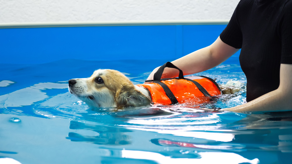
Physical therapies are an important part of good management of joint disease and joint pain. The main goals with physiotherapy are to improve joint range of motion, improve muscle mass and tone, address atrophy, manage pain and to improve stability. Physical therapies can include manual therapy, electrotherapy, acupuncture, physiotherapy and hydrotherapy. It is important to discuss the length of treatment and therefore the potential cost with owners before starting on this course.
Exercise and lifestyle adaptation
Exercise management or modification should be discussed with owners to make sure that animals receive the appropriate amount of exercise for their condition, their pain levels and their muscle mass and tone. Owners may feel like they are being good owners by providing lots of exercise but may actually be doing too much exercise rather than too little. There is no value in exercising beyond the point of fatigue, so if owners are starting to see hindquarters dropping, or more pronounced scuffing or lameness, this often is a sign that the tissues have fatigued.
It is important to consider the type of surface being exercised on, warm up and cool down periods, and the type of harness being used.
Weight management
Nurses and practices will be familiar with running weight clinics but these are critical in this context. Without improving the body condition score of an obese animal, it is very difficult to obtain the improvements that would be expected with the other therapies.
Joint supplements
There is evidence that omega acids have an anti-inflammatory effect when used at the correct ratio, but little evidence for changes in vivo from taking glucosamine chondroitin. Anecdotally, people say that glucosamine chondroitin can improve joint pain, so it is unlikely to do any harm in a well recognised supplement, but generally the omega acid content of any supplement is more important.
This workshop can be watched on demand at https://www.vetnurseworkshops.co.uk


