
In the author's veterinary practice negative pressure wound therapy (NPWT) is used in a variety of wound care cases. Publications on the technique date back 16 years, however, only minor changes have been made to the unit itself since it was first used: the unit has been made smaller; there is increased safety due to unit alarming; changes in the dressing associated and the ability for the unit to remove much more fluid (Murphy and Evans, 2012).
Large, contaminated wounds lend themselves to management with NPWT, along with dehiscing surgical wounds (Murphy and Evans, 2012); degloving injuries and skin avulsions are also indications for use along with chronic non-healing wounds. The fluid that is removed during NPWT contains high levels of proteolytic enzymes which if not removed can result in a non-healing wound environment. Less commonly compartment syndromes, for example abdominal or myofascial, are indications for NPWT management (Nolf and Meye-Lindenberg, 2016). Enterocutaneous fistulas now, despite early reservations, respond well to NPWT, and orthopaedic traumatic wounds benefit from this method as granulation tissue is evident on hardware, grafts and meshes (Murphy and Evans, 2012). Best (2014) reported the use of the unit in a slow healing distal extremity — the negative pressure applied to the wound lowers the interstitial pressure, lower than the capillary pressure, which in turn allows the capillaries to reopen and blood flow to the wound to increase.
The use of NPWT in wound management has increased in recent years as veterinary patients are now being treated for increasingly complex wounds, where veterinary professionals strive for a decreased treatment time, and to help avoid multidrug resistance and complications which can prolong wound closure (Nolf and Meye-Lindenberg 2016). Healing times have been reported to have been halved using NPWT compared with using a deficit foam dressing or traditional bandaging (Nolf et al, 2015). This method also lends itself to a decrease in number of dressing changes due to faster healing as a result of a quicker reduction in wound depth (Upton et al, 2013).
This management technique also extends to prevention of postoperative seroma formation or oedema (Perry and Whitte, 2013a). NPWT has been used to aid the ‘take’ of skin grafts enhancing re-epithelisation. The therapy encourages good communication between the graft and the wound bed, the unit effectively suctioning the graft down onto the site and providing an active drain to the area (Best, 2014; Nolf, 2015).
NPWT results in a much reduced size of wound, increased viability of skin grafts, improved response to antibiosis and reduced time of requirement for open wound management (Upton et al, 2013). The reduction in incidence of biofilm associated infections is also documented with early application of the method, which can be extremely challenging (Goldsworthy and Mcalinden, 2011). This equates to fewer bandage changes, less risk of hospital acquired infections due to increased length of stay, and reduced cost for the client (Perry and Witte, 2013b).
NPWT is seen to be most effective in the proliferative stage of wound healing, although it can be applied at any of the healing stages where appropriate debridement has occurred (Putnis et al 2014). Debridement is key to reducing the incidence of bacterial growth and to help encourage proliferation of granulation tissue (Howe, 2015). Inadequate debridement can lead to necrotic tissue and abscess formation. Therefore it is essential that any devitalised tissue is removed before the NPWT is applied (Schintler, 2012).
How it works
NPWT promotes wound healing by delivering negative pressure (a vacuum) at a wound site through an applied dressing. The wound edges are drawn together (Goldsworthy and Mcalinden, 2011), any infectious material is removed and granulation tissue is encouraged to form at a cellular level (Perry et al, 2015). There are numerous wounds that can have this method applied:
The V.A.C.® Therapy System (KCI, 2016) which is currently used in the author's practice, comprises three essential components that actively work together to help promote wound healing through granulation tissue formation.
The V.A.C.® Therapy Unit provides intermittent and continuous therapy with integrated patient safety features. The unit will alarm if there is any change in pressure indicating a leak or blockage and will also alarm if the battery is low. The collection chamber when full of fluid will also initiate an alarm for easy use by personnel (KCI, 2016). T.R.A.C.™ (Therapeutic Regulated Accurate Care) Pad Technology connects the wound to the unit to allow the negative pressure. It will regulate pressure at the wound site to provide accurate delivery of prescribed therapy settings. The pad will monitor and maintain target pressure at the wound site for consistent therapy delivery. It will reduce tubing blocks and false alarms through enhanced fluid dynamics (KCI, 2016). The V.A.C.® GranuFoam™ Dressings, which are placed directly into the wound cavity, are either a thick silver foam or thinner white foam. They help provide the necessary mechanisms to promote granulation tissue formation.
Under negative pressure, V.A.C.® Therapy Unit with proprietary V.A.C.® GranuFoam™ Dressing will apply mechanical forces to the wound to create an environment that promotes wound healing. These forces are known as macrostrain and microstrain (KCI, 2016).
The vacuum generated causes a three-dimensional contraction of the wound, bringing the wound edges together known as macro-deformation, or the mechanical creep effect or macrostrain (Perry and Witte, 2013a). Macrostrain is this visible change that occurs when negative pressure contracts the foam that is in direct contact with the wound bed, and draws the wound edges together (Putnis et al, 2014). There is evenly distributed negative pressure over the wound bed, and this allows removal of exudate, any infectious materials, and reduces oedema (KCI, 2016).
The wound is stabilised against any thermal and oncotic fluctuations which therefore prevents fluid evaporating and any scab formation, keeping the wound bed moist, which is critical for the enzymatic processes required for healing (Perry and Witte, 2013a). The dressing applied will also protect the wound from any mechanical disturbance (KCI, 2016). The vacuum will also decrease the interstitial pressure, and as a result the wound oedema subsequently decreases and any fluid is transported away from the wound (Putnis et al, 2014). Due to the interstitial pressure gradient, a mechanical deformation of the fibroblasts and collagen network occurs (Perry and Witte 2013a). The cells at the tissue/foam border undergo micro-deformation or microstrain which leads to cell stretch. Microstrain promotes perfusion of tissue cells. This in turn promotes granulation tissue formation as cell migration can occur and cell proliferation (KCI, 2016).
Problems encountered with wounds seen in veterinary practice and how the NPWT can aid wound healing are shown in Table 1.
| Problems for wound healing | Mechanism |
|---|---|
| Bacterial burden | Removes infectious materials |
| Inadequate protection against infection | Provides protected wound healing environment |
| Excess exudate | Removes exudate |
| Excess oedema (interstitial fluid) | Reduces oedema (interstitial fluid) |
| Absence of moisture | Provides a moist wound healing environment |
| Lack of adequate blood flow | Promotes perfusion |
| Lack of granulation tissue formation | Promotes granulation tissue formation and draws wound edges together |
Application
Before the NPWT is applied, the wound requires debridment and lavage. A generous clip is required to allow both aseptic technique and for sufficient clipped skin for the dressings to adhere to. A sterile skin prep should then be applied to the skin, such as 2% povidine iodine (chlorhexidine can be cell toxic), avoiding the wound itself. The wound itself can be flushed with saline or alternative products such as Prontosan (Braun) (Smith, 2013). This should be done in a preparation area prior to theatre.
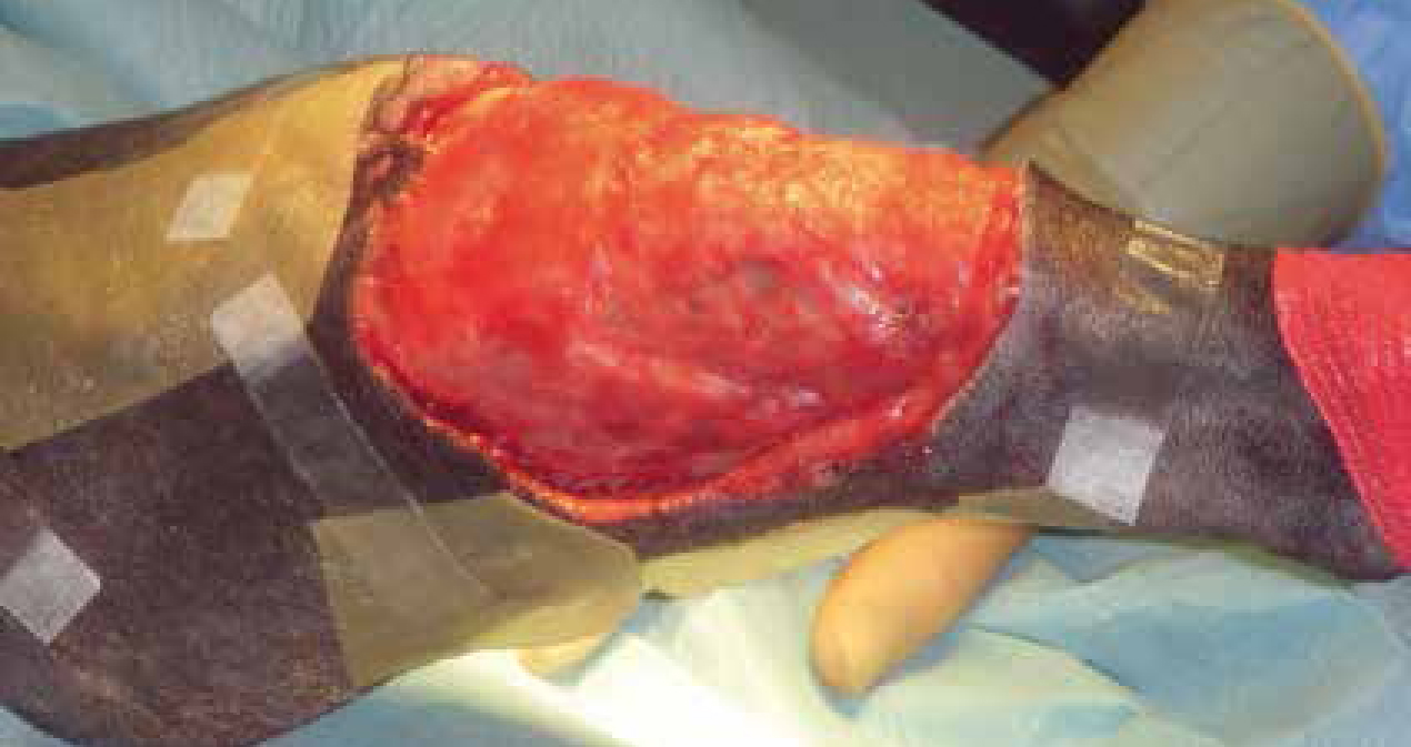
Once the patient has been transferred to theatre, the wound can be debrided by the veterinary surgeon using aseptic technique. Following debridement, the skin should be dried using sterile swabs. A foam dressing should be cut to the size that will fill the deficit and placed within the wound. It is important to ensure good contact between the dressing and all areas of the wound. Foam dressings help provide the necessary mechanisms to promote granulation tissue formation. The deep wounds lend themselves to dressings that should fill the deficit, but should not overlap onto the skin so as not to cause irritation as shown in Figure 1. Foams with silver can be used if there is an increase in bacterial load (Stinner et al, 2011). For superficial wounds gauze can be used and also to bridge closed incisions that require therapy. Both foam and gauze should be of a grading of 400–600 µm (Perry and Witte, 2013b). If required, special gel stripes can be used to assist in sealing the area around the wound (Figure 2).
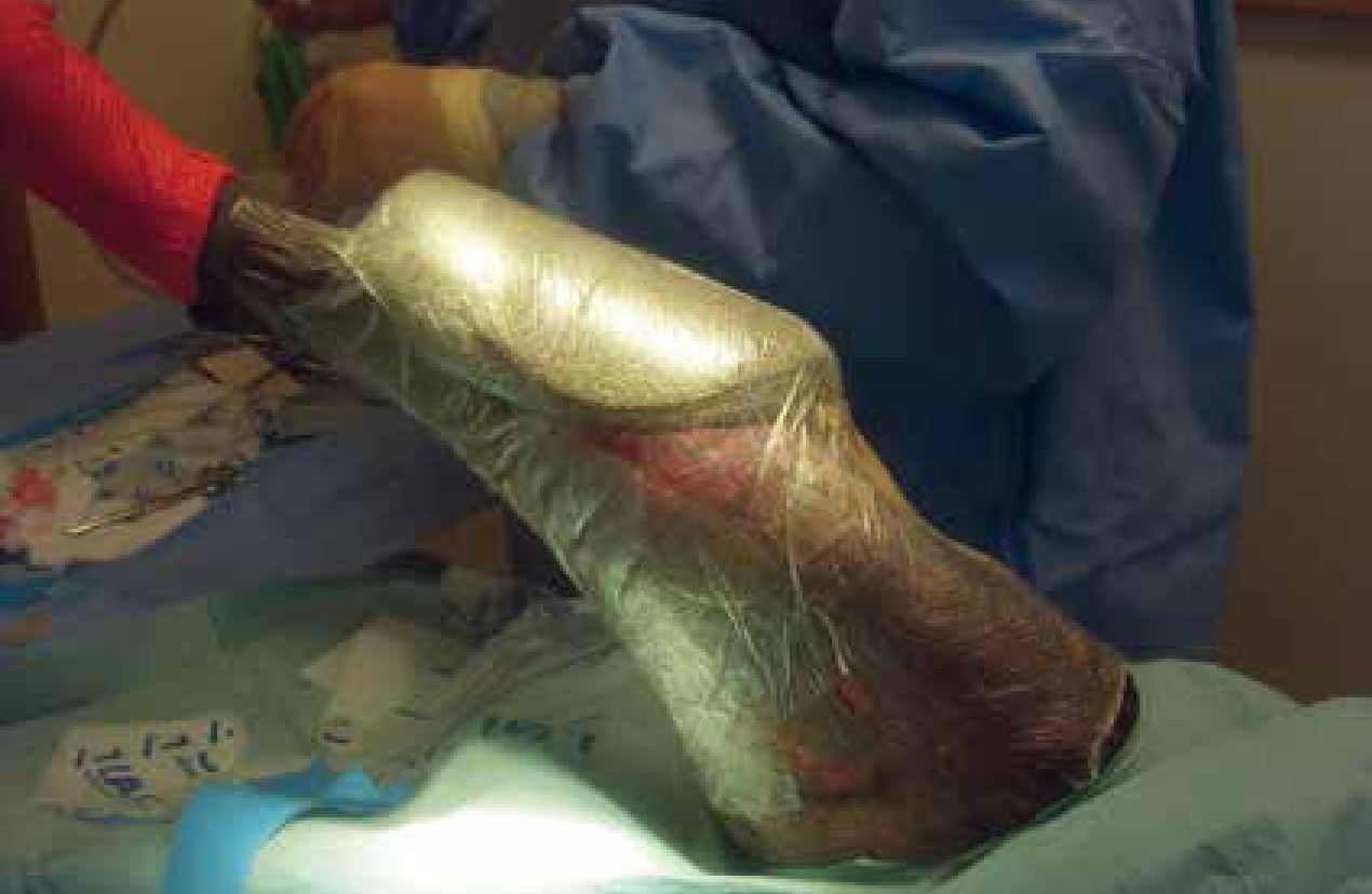
To ensure a good seal the area around the wound should be clipped, cleaned and dried, as discussed above. Occasionally, when the drape fails to adhere to the skin, an adhesive aerosol or liquid can be used to help adhere an occlusive dressing, for example V.A.C Drape®, over the area concerned. There should be a 3–5 cm clearance of the occlusive dressing around the foam dressing, which is placed within the wound, to ensure a seal (Perry and Witte, 2013b). This is best achieved by using small overlapping pieces of occlusive drape rather than trying to apply one big piece (Figure 3).
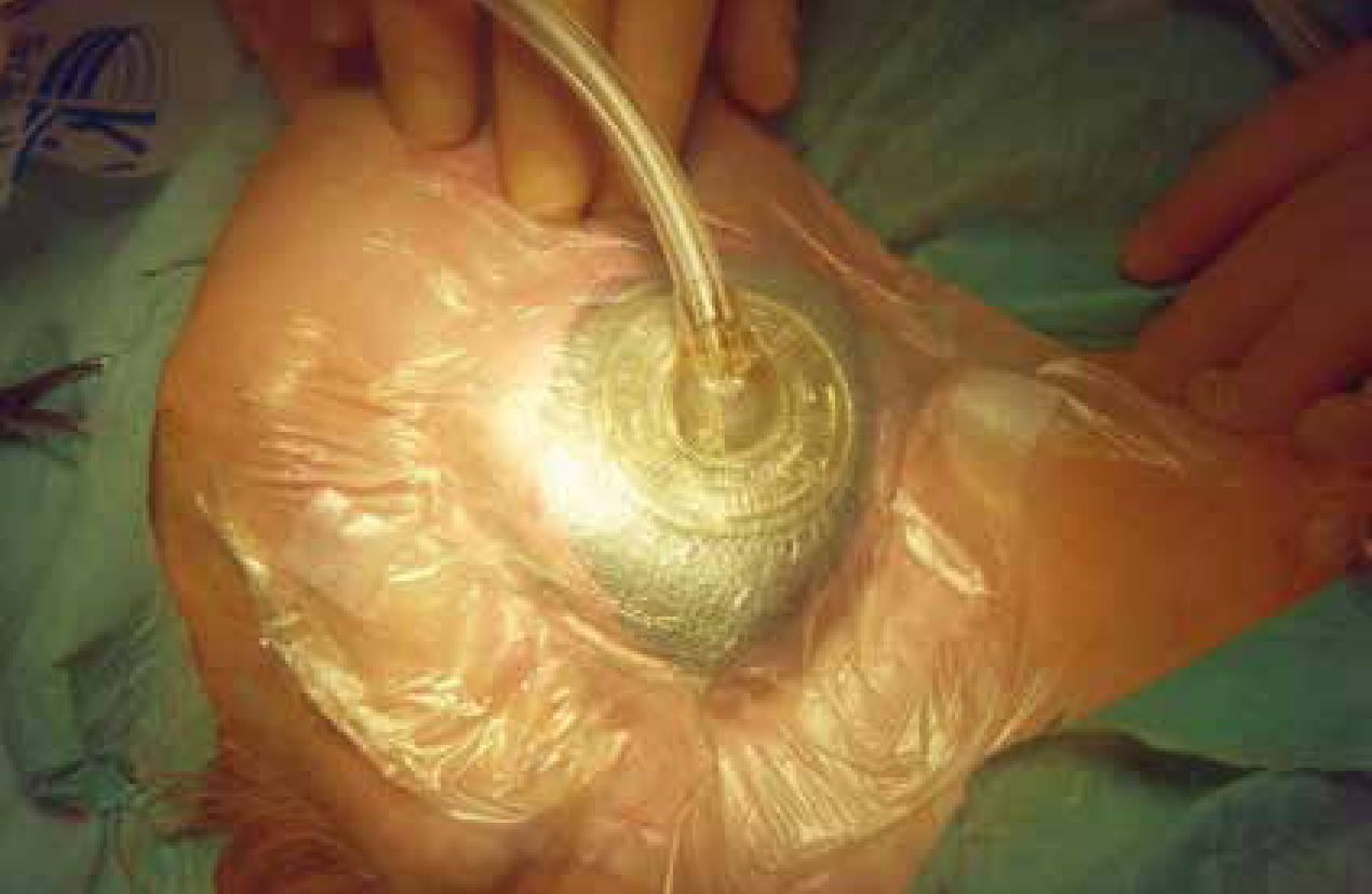
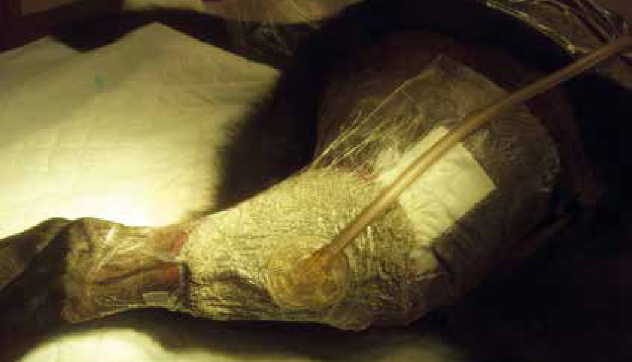
Careful application of the NPWT consumables is required to ensure airtight sealing of the area. Producing an airtight seal can be challenging (Perry and Witte, 2013b). A hole is cut into the drape centrally over the foam, and the pressure evacuation tube is applied to the dressings, to eliminate any fluid from the site; this initially contracts the foam known as the raisin effect (Perry and Witte, 2013b) when the vacuum device is switched on (Figure 4,5). It is important to guarantee that the therapy is not interrupted for longer than 2 hours per day. If the device is not properly working, bacteria within the wound are granted optimal conditions to proliferate, resulting in wound infection and tissue maceration (Goldsworthy and Mcalinden, 2011).
Considerations and complications
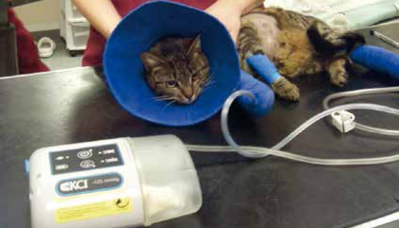
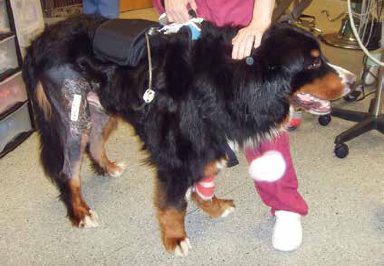
In veterinary medicine the common setting for NPWT is -125 mmHg. Stanley (2012) has documented that to diminish postoperative oedema, -50 to -75 mmHg can be used, but veterinary units available do not have the capabilities for alteration of pressure. The intermittent negative pressure will improve blood flow and granulation tissue formation, however it has been shown in some human studies to cause some discomfort (Morykwas et al, 1997). Schintler (2012) agreed with Stanley (2012) that a negative pressure of -125 mmHg encourages maximum formation of granulation tissue and will increase wound healing, and Perry and Witte (2013a) state that -125 mmHg can quadruple blood flow to the capillaries. As seen in Figure 5, patients tolerate the system well, even feline patients. Figure 6 shows a giant breed canine patient wearing a unit — like human patients larger dogs can carry them around with them.
Consideration should always be given to the nature of wound, the age of the patient, whether infection is present and the location of the wound as all these can have an effect on healing (Nolf et al, 2015), and can impact on how successful the NPWT treatment can be and how long the system is used for.
Bandage change frequency should be determined on a case by case basis, and quite often more frequent changes are required early in the wound process, normally to facilitate debridement (Perry and White, 2013a). Literature suggests the unit should not be left without change for more than 72 hours. In human medicine toxic shock syndrome has been reported as a result of small fragments of foam left within the wound when granulation tissue had grown into the dressing, and remained in the wound once the dressing was removed (Perry and Witte 2013b). Every other or every 3 days is documented as best practice (Stanley, 2012; Nolff and Meyer-Linderberg, 2016)
As loss of an airtight seal can cause wound maceration (Stanley, 2012) it is imperative that continuous negative pressure is maintained, and that all personnel involved are aware of the unit's mechanisms and procedure if the alarm is sounded.
Contraindications of use
Contraindications include use on wounds that have a known neoplastic involvement, as use within an area of local malignancy could seed neoplasia further (Howe, 2015). Haemorrhage may occur when using this method due to erosion of any exposed arteries or veins, so its use in patients with coagulopathies should be cautious.
Despite an increase in use of this wound management technique, there is still a requirement for more blind studies to be conducted due to gaps in evidence-based medicine in veterinary patients (Schintler, 2012).
Conclusion
NPWT is an excellent tool to assist in wound closure in the veterinary practice. Complicated or static wounds can be managed more effectively than with traditional wound care techniques to aid in quicker recovery of patients, with shorter stays in hospital, which can minimise the risk of acquiring a hospital infection and with reduced cost to clients. In summary NPWT increases perfusion to wounds, reduces oedema and interstitial tissue fluid, stimulates granulation tissue formation, promotes reverse tissue expansion and reduces bacterial colonisation. Advances in NPWT, which are being echoed from the human field into the veterinary market, will allow the treatment of veterinary patients to be improved as new techniques and products are evolving, more complex wounds in challenging areas are being investigated so that this therapy can be used to its full potential.