
Dental disease is the number one clinical problem seen in small animal practice, and thus oral pathology is exceedingly common in small animal patients (Lund et al, 1999). In addition, there is a very wide variety of pathologies that are encountered within the oral cavity. These conditions often cause significant pain and/or localised, regional and systemic infection (Niemiec, 2008). This text is a quick reference for veterinary nurses to identify the common oral pathologies seen in the dog and cat.
Importance of oral examination
The only way to reveal oral pathology is through a thorough examination. An oral examination should always begin in a conscious patient (Wiggs, 1997; Gorrel, 2008). If the patient (due to aggression or impatience) does not allow a thorough visible inspection, the next step is sedation. The position of evaluator should be comfortable. Appropriate lighting, close access to the head, and ideally an assistant who records the findings will improve the quality of the examination. In addition, an assistant is a significant asset in the photographic evaluation of the patient (Bellows, 2004). At this stage of examination particular attention is paid to the occlusion (head symmetry, number of the teeth, and their relationship), dentition and dental deposits are carefully measured and recorded, and regional lymph nodes palpated.
Anaesthesia allows comfortable access, but the relationship between the maxilla and mandible is not the same as when natural tension of the masticatory muscles is present so it is less reliable for e.g. orthodontic purposes (Gawor, 2013a). However, it is only possible to perform detailed oral/periodontal assessment, intraoral radiography and other diagnostic procedures, such as biopsy or impression taking, in an anaesthetised patient.
After sedation and induction, the patient should undergo all necessary preparations to provide safe anaesthesia. After safe anaesthesia is assured, the entire oral cavity must be systematically evaluated using both visual and tactile senses. Careful visual examination should be performed along with the periodontal evaluation. Salient findings include (but are not limited to): fractured, mobile, or intrinsically stained teeth, foreign bodies, tooth defects such are caries or tooth resorption, and oral masses. The authors recommend that the patient be placed in dorsal recumbency for this step, as it will improve visualisation (Huffman, 2010).
The oral structures must be visually assessed, palpated and examined with the use of a dental explorer (dedicated to hard tissues) and periodontal probe (used for soft tissues) and with the help of the mirror and retractor. The oral mucosa present at cheeks, lips, palate, pharynx, tongue, and sublingual area is evaluated for its integrity and colour. Veterinary nurses should take into account that some anaesthetic drugs (e.g. medetomidine) influence vascularity of mucosa and reduce inflammatory signalment (redness) (Murrell, 2007).
The periodontal evaluation should begin with determining the plaque, calculus, and gingivitis index. These key pieces of information are best noted prior to the dental cleaning. Following this, the periodontal status is measured. The only accurate method for detecting and measuring periodontal pockets is with a periodontal probe, as pockets are not always diagnosed by radiographs (Tetradis et al, 2006; Niemiec, 2010).
Radiographic evaluation of oral structures is a mandatory and complementary part of oral examination. The value of radiographic evaluation in veterinary patients was proven in studies which found 27.8% of clinically important lesions in dogs and 41.7% in cats would be missed without full mouth radiography (Verstraete, 1998a; Verstraete, 1998b).
There are numerous books describing possible oral dental and maxillofacial diseases. Some of them are listed at the end do text as further reading. The oral problems presented below are statistically quite common and it is important to diagnose these conditions at the primary health care or general practice level. Despite the fact that they may lead to serious complications, the affected patients rarely display their discomfort, and behave almost normally. For the vast majority of pet owners, and unfortunately for a significant part of veterinary community (including nurses), until the patient stops eating, drinking or playing, the oral disease is largely ignored.
It is the authors' intention to accurately describe the selected common oral pathologies, their influence on oral, regional, and systemic health, and the necessity to manage them. Increased awareness of oral problems will increase dental revenue and improve the welfare of the veterinary patients. Periodontal disease requires a separate chapter and therefore was not a part of this article.
Persistent deciduous teeth
A deciduous tooth is considered persistent as soon as the permanent tooth erupts. There should never be two teeth of the same type in the same place at the same time (Niemiec, 2010).
Persistent deciduous teeth (Figure 1) are exceedingly common, especially in small and toy breed dogs. However, they can occur in any breed as well as cats. They create both orthodontic and periodontal problems if not treated promptly. It was previously believed that the persistent deciduous tooth caused the permanent tooth to become malocclused. However, it is now known that the permanent tooth erupting incorrectly causes the deciduous tooth to be persistent (Hale, 2005). Further, orthodontic problems begin within 2 weeks of the permanent canines starting to erupt. This is due to the deciduous tooth being in the place that the adult tooth wishes to occupy.
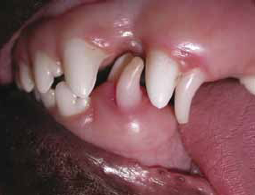
The periodontal issues occur due to a disruption of the normal maturation of the periodontium. When there is a persistent deciduous tooth, one area of the periodontium is not attached to the permanent tooth, therefore the periodontal attachment in that location will not be normal. It has been reported that the damage begins within 48 hours of the permanent teeth starting to erupt! This means that the adult tooth does not need to be completely erupted for these problems to occur. Thus, the deciduous tooth should be extracted as early as possible — do not wait until 6 months of age to perform the extractions along with neutering. In fact, the authors recommend that the owners of breeds prone to retain their deciduous teeth be instructed to watch for eruption of the permanent teeth and to present the patient for therapy as soon as this occurs.
Supernumerary teeth
This occurs when there are extra teeth in the arcade. In some breeds there is reported predisposition for supernumerary dentition (boxers, bulldogs) (Gawor, 2013b). If there is room for these teeth and they are not causing undue crowding or occlusal trauma, no therapy is necessary. If they result in crowding, selective extractions are recommended early to relieve the crowding and allow for natural cleaning ability (Holmstrom, 2013).
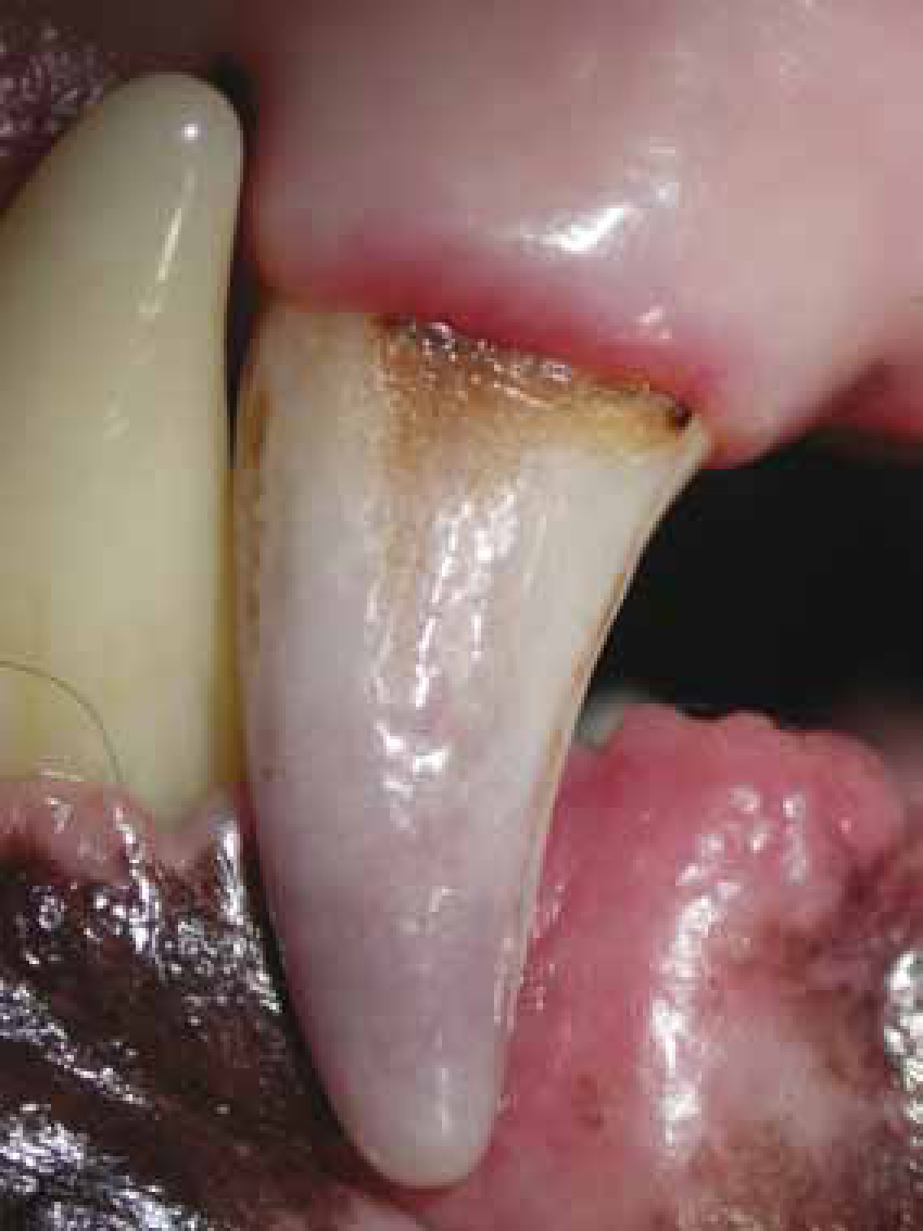
Intrinsically stained (discoloured) teeth
These teeth can appear pink, purple, yellow, or grey (Figure 2). The most common intrinsic stain seen in dogs is caused by pulp haemorrhage due to trauma. Tetracycline that is taken during tooth development causes dark discolouration (DuPont, 2010). Endodontic disease is also manifested by intrinsic staining. Non-vital teeth lose their natural defence ability and are often infected via the bloodstream, which is known as anachorisis. A study by Hale showed that only 40% of intrinsically stained teeth had radiographic signs of endodontic disease, however 92.7% are non vital. Therefore, do not rely on radiographic appearance to determine vitality; all discoloured teeth should be definitively treated via root canal therapy or extraction (Hale, 2001).
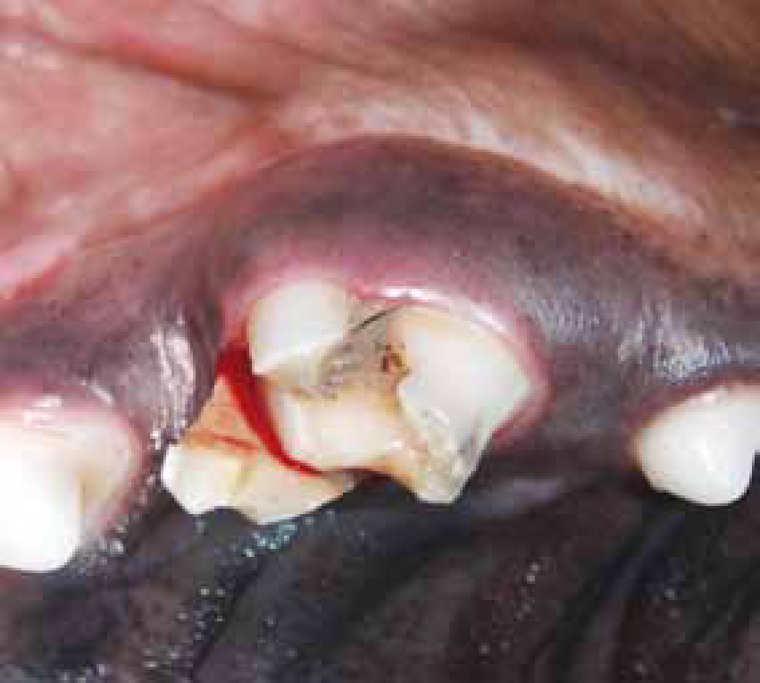
Fractured teeth
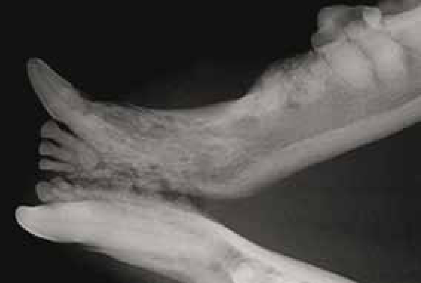
The two main types of crown fracture seen in veterinary medicine are complicated and uncomplicated. Complicated fractures occur when the endodontic system (pulp) is directly exposed (Figure 3). Uncomplicated fractures are when the damage is limited to the enamel +/− dentin (Figure 4). Both types require therapy, however treatment for each is often different.

Uncomplicated crown fractures are a very common finding on oral examination, particularly in large breed dogs. These fractures result in dentin exposure which creates dentinal sensitivity via the exposed tubules resulting in significant pain for the patient. In addition, some of these teeth will become non vital due to the traumatic incident, pulpal inflammation, or bacterial invasion via the dentinal tubules. For these reasons, it is recommended that these teeth be radiographed to ensure vitality. If the teeth are non-vital (which is evidenced by periapical rarefaction or a wider root canal than contralateral teeth) endodontic or exodontic therapy is required. If the teeth appear vital, the application of a bonded composite is recommended to decrease sensitivity (Niemiec, 2005). The complicated tooth fracture should receive specialist consultation and treatment.
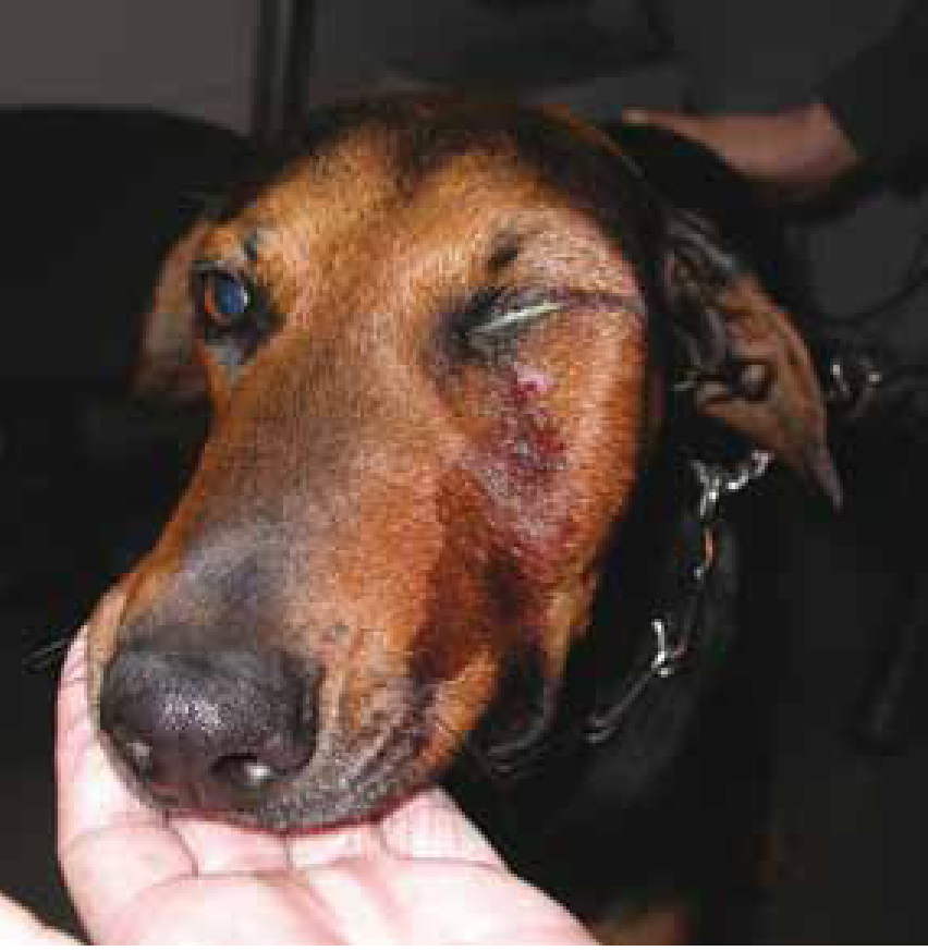
Abscessed teeth
These are also commonly called ‘carnassial abscesses’ because they most often occur with fractures of the maxillary fourth premolar. However, any infected tooth can result in a clinical abscess (Figure 5). It is critical to note that these teeth have been dead and infected for a long time (sometimes years) and have just recently demonstrated outward clinical signs. The patient has been subclinically infected for a long time. Tooth root abscesses should be a differential diagnosis in any case of acute facial swelling. They are typically associated with a complicated crown fracture. However, it is critical to note that uncomplicated crown fractures can often result in clinical abscessation.
Antibiotics can be used to temporarily resolve the pain/infection/and swelling. However, once the tooth becomes infected, there is no way to effectively medicate the root canal system. Further therapy is therefore required, regardless of the resolution of the acute problem. The infection will smoulder for a while and then reabscess, leaving the patient to suffer through that entire intervening period.
Definitive treatment options include complete extraction or ideally root canal therapy. Furthermore, the prognosis with root canal therapy of an abscessed tooth is very similar to this procedure on a vital tooth (Niemiec, 2005).
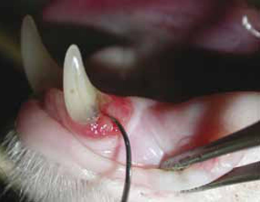
Luxated/avulsed teeth
A luxated or avulsed tooth has been traumatically torn out of the alveolus. This most commonly occurs with the canine teeth (especially maxillary), but incisors can be affected as well. This typically occurs following dog fights, but can also result from significant cage chewing or trauma. These cases should be referred to a veterinary dentist as soon as possible for replacement and stabilisation. This is one of the true dental emergencies, as saving these teeth is very time dependent. The fixation method is typically a figure-8 wire and acrylic splint, however some veterinary dentists prefer a flexible fixation to avoid ankylosis and root resorption. At this time, or at time of splint removal, a root canal will be necessary on the affected tooth (Holmstrom, 2013).
Tooth resorption (TRs)
TRs are a very common malady in cats but have also been reported in other species (Figures 6a and 6b). Reports vary as to their incidence, but approximately 60% of cats over 6 years of age have at least one, and those that have one typically have more. Pain and malfunction of the digestive apparatus are the major signalments of the disease. TR is very often associated with caudal stomatitis in cats (DuPont, 2010).
It used to be thought these lesions were caused by odontoclasts which are cells that are responsible for the normal remodeling of tooth structure, however other types of cells, e.g. cementoblasts or osteoblasts, have also been found to be involved in this disease. These cells are activated and do not down regulate, resulting in tooth destruction.
Gingival inflammation is one sign of the resorptive process, therefore any redness of gingival margins should be noted, evaluated, and radiographed.
There are currently two recognised forms of feline resorptive lesions, type 1 and type 2. An additional type is regarded as presence of the two first types within same tooth. Clinically, they appear very similar, as dental defects that are first noted at the gingival margin. However, advanced cases will show significant tooth destruction and may appear to be a fractured tooth. The best diagnostic tool for differentiating between types of felines resporptive lesions is dental radiology; with type 1 lesions, there is no replacement of the lost root structure by bone, whereas with type 2 there is generally marked replacement of the lost tooth structure (Reiter et al, 2005).
Recently, crown amputation has been developed as a treatment option for advanced type 2 lesions as it results in significantly less trauma and faster healing than complete extraction. This procedure, although widely accepted, is still controversial. Most veterinary dentists employ this technique, however in widely varying frequency. Veterinary dentists typically employ this treatment option only when there is significant or complete root replacement by bone. Unfortunately, the majority of general practitioners use this technique far too often — crown amputation should only be performed on teeth with radiographically confirmed advanced type 2 TRs which show no peri-apical or periodontal bone loss. Crown amputation should not be performed on teeth with: type 1 TRs, radiographic or clinical evidence of endodontic or periodontal pathology, inflammation, or infection; or in patients with L/P stomatitis. Those practitioners without dental radiology capability should not perform crown amputation. In these cases, the teeth should either be fully extracted or the patient referred to a facility with dental radiology (DuPont, 2010). Studies show preliminary results of conservative treatment of teeth resorption to be promising (Mohn et al, 2009), however filling the cavities does not provide good prognosis presumably because of complex and not yet clarified aetiology (DuPont, 2005).
Missing teeth
There are several reasons that teeth may be missing. These reasons include: congenitally missing, previously extracted or exfoliated, fractured (or extracted) with retained roots, or impacted. The first two scenarios do not require therapy, whereas the latter two typically necessitate intervention. Therefore, dental radiographs are indicated in all cases of ‘missing teeth’.
If dental radiographs reveal retained roots and evidence of inflammation or infection (clinical or radiographic), the teeth should be surgically extracted. If they are ‘quiet’, the owners should be informed and given the option of having the teeth extracted.
Impacted teeth are defined as any tooth that has not erupted by its normal time. This is generally considered to be the time when the surrounding or contralateral teeth have already erupted. The most common cause of impaction is the presence of an overlying structure that interferes with normal eruption. Unerupted teeth always have the potential for odontogenic cyst formation and require either extraction or continuous monitoring (Lobprise and Wiggs, 1992).
Oral neoplasia
The oral cavity is the fourth most common place to encounter neoplastic growths (Figure 7). In dogs a large proportion of proliferations are reactive or benign, while in cats the majority of proliferations are malignant (Harvey and Emily, 1993).
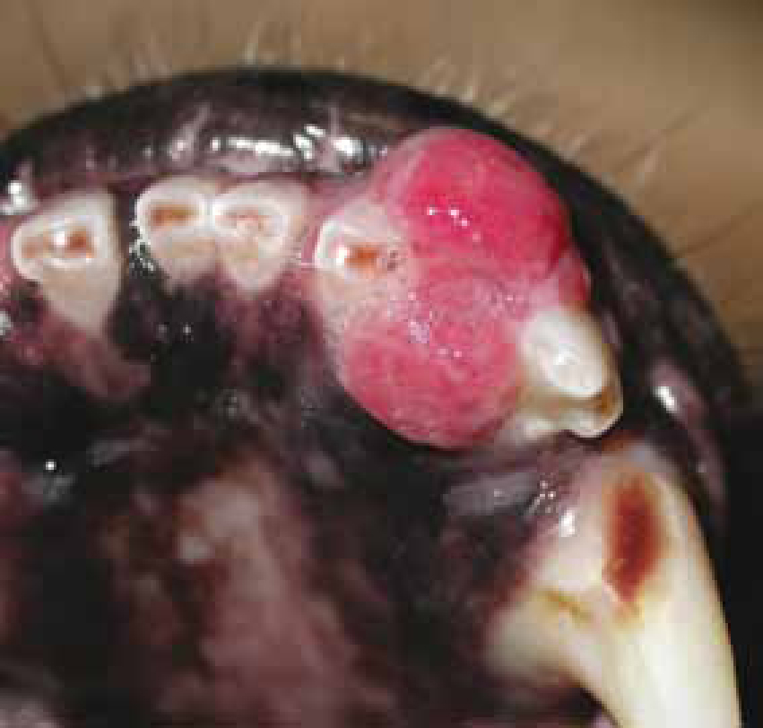
Any growth, proliferative, or abnormal non-healing lesion in mouth must be biopsied. The purpose is to diagnose the potential neoplastic process at an early stage (Dhaliwal, 2010).
The most common canine oral growths are the epulids which are any localised, exophytic swelling on gingiva (Verstreate, 1999). Approximately half of the epulides are reactive lesions but still a large proportion can be locally aggressive or neoplastic lesions. Therefore biopsy is always mandatory (Verstreate et al, 1992).
Neoplastic lesions are classified into two groups: the odontogenic and non-odontogenic tumors. The first group contains odontomas, ameloblastomas, and peripheral odontogenic fibromas. In the second one, malignant melanoma is reported to be the number one oral malignancy in dogs (30–40%) and squamous cell carcinoma the most common in cats (60–70%). Fibrosarcoma is less common in dogs and is a distant second in cats. Osteosarcoma is mainly diagnosed in large breed dogs (Verheart, 2010).
Melanomas are typically seen in older dark pigmented dogs, and are not only locally aggressive; they also metastasise very early in the course of the disease (Holmstrom, 2013). A combination of aggressive surgery, radiation therapy, and chemotherapy is the best way to treat this disease process. In addition, a vaccine has become available recently that shows promise as an adjunct therapy for this disease process (Bergman et al, 2003).
By far the most common malignant oral tumor in cats is a squamous cell sarcoma. Fibrosarcomas are a distant second. Both of these tumors are typically seen in older cats, are locally aggressive, and are late to metastasise. The only therapeutic option at this point is early, aggressive surgery (2 cm surgical margins).
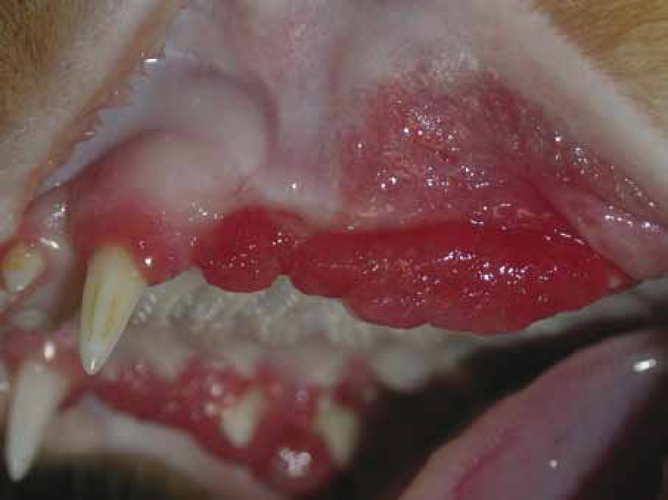
Feline caudal stomatitis
Feline caudal stomatitis (Figure 8) is a relatively recently discovered disease process seen in cats which is currently very frustrating to treat. While the true etiology is currently unknown, the best description is a severe immune-mediated reaction to dental tissues. Some feel that this may actually be a group of disease processes that look the same clinically, which is why they can be very frustrating to treat. Although there is suspected breed predilection for this condition (Siamese and Burmes) it is diagnosed in all cats (Lyon, 2005).
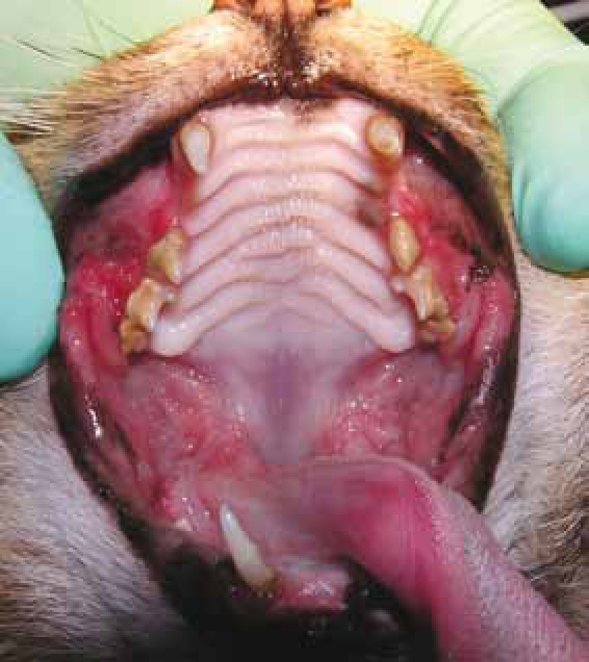
The history will generally include anorexia, drooling, gagging, and pain during mastication. Physical examination will typically include a thin pet with unkempt fur. The oral examination will reveal severe stomatitis throughout the gingiva. The inflammation will most commonly be worse on cheek teeth than canines and incisors. However, caudal mucositis is the key clinical finding. Severe hyperplastic inflammation to the gingiva can result from periodontal disease; however caudal mucositis will not be present.
A pre-operative blood panel will generally show a marked elevation in globulins (polyclonal gammopathy) and total protein (DeBowes, 2010). Histopathology is recommended but not required. There have been a few cases seen by the authors with presentations suggesting another pathology (fungal, immune mediated). In this case full mouth extractions would be ineffective (see below).
Extraction is currently the only effective long-term treatment for this disease process in cats. In the authors' experience, the sooner this is done, the better the pets do both post operatively as well as long term. For extractions to be successful, the teeth must be completely removed (Gorrel, 2008). Therefore post-operative radiographic confirmation of complete extraction of the tooth roots is recommended. Following the confirmation of complete removal of the teeth, aveloplasty should be performed to remove the periodontal ligament, and smooth any rough bony edges. This is typically performed with a coarse diamond bur. Studies report a 60% success rate when all teeth caudal to the canines are extracted (Hennet, 1997); however the authors' experience has not been as good. Anecdotally, whole mouth extractions have a success rate of approximately 90–95% for clinical remission.
Feline juvenile (puberty) gingivitis/periodontitis
Juvenile periodontal disease (Figure 9) is inflammation that occurs soon after permanent tooth eruption. This syndrome can be separated into two categories: feline hyperplastic gingivitis (inflammation is confined to the gingiva); and juvenile onset periodontitis. At the time of writing, the etiology of this condition remains unknown. However, in humans there is a period of increased susceptibility to gingivitis during the pubertal period (puberty gingivitis). A genetic predisposition has been reported in Siamese, Somali, and Maine Coon cats (Niemiec, 2011).
Hyperplastic gingivitis appears as gingival enlargement and significant inflammation which is confined to the gingiva and begins during the eruptive period of the permanent dentition. Bleeding during mastication and on oral examination are common findings.
In the management of both of these conditions, early (9 months of age) and frequent (every 6–9 months) dental prophylaxis (even if only minimal plaque is present) along with strict home care is critical to decrease inflammation. Ideally, home care consists of daily brushing, as it is the gold standard for plaque control. Other home care alternatives include chlorhexidine rinses as well as plaque control diets and treats. In cases where gingival hyperplasia is present, early gingivectomy is recommended to remove psuedopockets, decrease inflammation, and facilitate plaque control (both professional and home care). Finally, extraction of any significantly diseased teeth is warranted to decrease the degree of inflammation (Hale, 2005). If properly treated, disease in approximately 50% of these cats will resolve, the other 50% will have a lifelong struggle.
Immune-mediated diseases affecting the oral cavity
These diseases are caused by an inappropriate response of the immune system resulting in tissue destruction by its component cells. Immune-mediated disease has been divided into two distinct types. The first is primary immune mediated, also called ‘autoimmune’, in which the antibodies are directed against normal body tissues. In secondary immune-mediated disease, simply termed ‘immune mediated’, the destruction is created by a reaction other than normal self antigens.
In primary (autoimmune) disease, antibodies develop against normal body tissues and induce lesions by passive transfer. Secondary (immune-mediated) disease is caused by antigens that are foreign to the patient. In general, these are drugs, bacteria, or viruses that create an immunologic response. Antibiotics are frequently implicated in secondary immune-mediated disease, as are numerous other medications. Parasiticides have also been implicated in immune-mediated disease. Finally, secondary cases have been reported associated with malignancies (Niemiec, 2010c).
The classic therapy for the primary form, or autoimmune disease (pemphigus complex, bullous pemphigoid and SLE) is immunomodulation. This is typically achieved with corticosteroids. Many patients with predominantly oral lesions respond favorably to immunosuppressive corticosteroid doses (2 to 3 mg/kg twice daily for 10 days) tapering with response. It must be noted that at these doses, severe side effects (including death) are quite common. For this reason, it is recommended to add ‘steroid sparing agents’, such as azathioprine, in order to decrease the dose of steroids and the incidence of steroid-induced side effects, especially in chronic cases.
Conclusion
Oral diseases are the most common medical problem in dogs and cats and they are very often missed or misdiagnosed. The only way to improve this situation is veterinary team education and disciplined performance of an oral examination along with every overall physical examination. Every patient must receive an oral examination on each visit to the hospital. Most oral pathologies cause related pain and/or infection. It is the goal of veterinary team to relieve these conditions. If the clinical finding requires therapy beyond the skills and equipment of the practice, a referral to a veterinary dental specialist should be made. Every effort should be undertaken to properly diagnose and treat oral and dental maladies.