
Infections transmitted by ticks are increasingly recognised as important causes of disease in dogs. Canine protozoal, rickettsial and bacterial tick-borne diseases, such as babesiosis, borreliosis, anaplasmosis, ehrlichiosis, are some of the diseases that have been described with increasing frequency throughout the world in recent years (Githeko, 2000). Better animal care, better diagnostic tools used by microbiologists and parasitologists, the curiosity of veterinarians faced with unusual clinical syndromes in their patients, and a broader geographic distribution of the tick vector through pet travels, translocation or commercial trade of pet dogs, are some of the factors contributing to the emergence and increased recognition of these, and potentially other, tick-borne diseases (TBDs).
In recent years, the geographic range of many of TBDs has expanded and several novel infections have been described, suggesting that the range of pathogens transmitted by ticks is more extensive than previously assumed. Babesia and other tick-borne pathogens share a common link, which is tick transmission. Specific tick species preferentially transmit different pathogens, dogs can be sequentially or simultaneously infested with more than one tick species, and a single tick can transmit more than one pathogen leading to co-infections. Both the tick species and the pathogens that they transmit can vary substantially within and between various geographic regions. These factors make the diagnosis and medical management of TBDs a complex and challenging task for veterinary professionals.
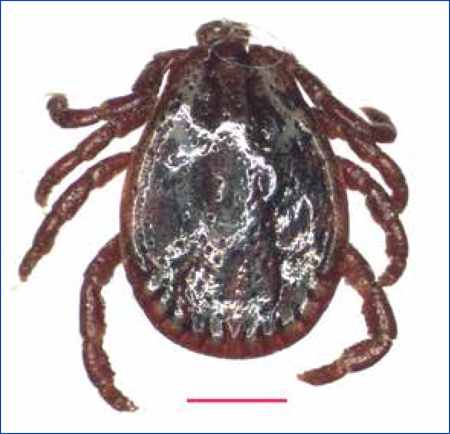
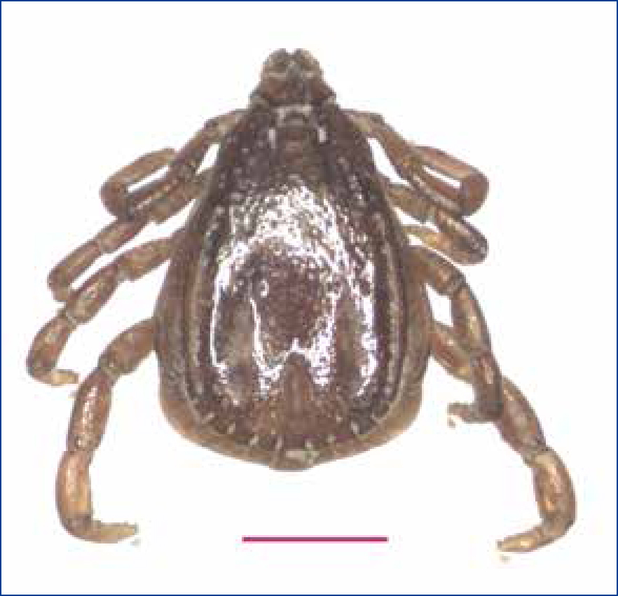
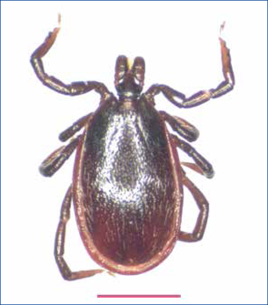
As a result, understanding of tick distribution and species abundance in the UK is more important than ever. This was the motivation for the launch of the first comprehensive survey of ticks in dogs, the ‘Big Tick Project’, which was carried out by Professor Richard Wall from the University of Bristol (Abdullah et al, 2016). This project was conducted over a 16 week period (April–July 2015) and involved voluntary collections of ticks by veterinary practitioners from 1094 veterinary practices across the UK. Of the 6555 tick samples collected from infested dogs, Ixodes ricinus (Figure 1) was identified on 5265 dogs (89 %), Ixodes hexagonus on 577 (9.8 %) and Ixodes canisuga on 46 (0.8 %). Ten dogs had Dermacentor reticulatus (Figure 2), one had Dermacentor variabilis, three had Haemaphysalis punctata and 13 had Rhipicephalus sanguineus (Figure 3). All the R. sanguineus and the single D. variabilis were on dogs with a recent history of travel outside the UK. These tick species represent significant risks to dogs, other pets and humans for a range of TBDs.
How serious can the problem of exotic infections introduced by ticks be? The immediate answer came when the first cases of babesiosis in dogs were reported in the UK in early 2016. Worryingly, none of the dogs that contracted the disease had travelled out of the UK. Even though these cases are confined to Harlow in Essex animal disease experts warned that the spread of the disease to the rest of the UK is inevitable due to the relaxation of the pet travel rules (Phipps et al, 2016). It used to be compulsory for imported dogs to be treated for ticks before entering the UK and Ireland, but the requirement was dropped in January 2012 to comply with EU regulations (Abbott et al, 2011).
The next sections will discuss the crucial roles that veterinary nurses can play in supporting pet owners in recognising and handling ticks and TBDs followed by a brief account of the major diseases ticks can transmit, and which can have a significant impact on the health and welfare of dogs in the UK.
Tick bite prevention
Advising clients about prevention of tick bites, especially in the spring and summer months, may help prevent exposure to some dangerous TBDs. Some effective ways to avoid tick bites are to wear protective clothing with long sleeves and long trousers tucked into socks or boots, and treat socks and trouser legs with a permethrin-containing insecticide. It is worth noting here that there is a risk to animal owners from TBD given they are likely to have been in the same environment as the dog. Hence, pet owners should check pets for ticks after walks. If one is found on the body it should be removed completely. The body of the tick should be grasped gently and vertical traction applied, using blunt, medium-tipped, angled forceps, until it displaces. Commercially available devices (known as tick twisters) especially designed for tick removal using ‘twist and pull’ action can also be used. Tick removal methods that should be avoided include applying a hot match to the tick body, covering the tick with petroleum jelly, nail polish, alcohol, or gasoline, using lidocaine, and passing a needle through the tick. When an improper technique is applied, parts of the mouth may remain in the skin, which can lead to infection or elicit inflammation. If pet owners spot signs in their dog, such as weakness, pale gums or ‘coffee-coloured’ urine, then they should contact their veterinarian immediately. If no signs follow exposure to tick bites, empiric treatment is not generally indicated. But, in animals with clinical findings suggestive of a TBD, treatment should not be delayed for laboratory confirmation.
People sometimes only discover that their dog had been bitten by ticks when clinical signs of a TBD appear. By then, treatment is expensive, if even treatable. Thus, it is essential to advise clients to use acaracide products routinely and year round to prevent tick-borne infections.
Because 24 to 48 hours (or even less duration in Rickettsias and Ehrlichia) of attachment to the host are required for infection to occur, early removal can help reduce transmission of infection and prevent disease. The same tick may harbor different infectious pathogens and transmit several with one bite. Hence, identifying the tick species is clinically helpful because it will alert the veterinarian to the diseases that may have been transmitted. Such information must be obtained quickly. The collected ticks should be morphologically identified using identification keys available for local species (e.g., http://bristoltickid.blogs.ilrt.org). Also, dog owners and members of the veterinary profession can send any ticks they might find to Public Health England's Tick Recording Scheme or the Big Tick Project (http://www.bigtickproject.co.uk) for species identification. However, morphological identification can be difficult because it requires some entomological expertise and any specimen that is damaged or immature is difficult to identify. Ticks removed should be effectively disposed of (https://www.gov.uk/guidance/tick-surveillance-scheme) to prevent contamination of the environment.
Travelling pets
Veterinary nurses often encounter clients preparing to travel to potentially tickor TBD-endemic regions. They should have basic knowledge of pathogen transmission and tick prevention strategies. The risk of TBD acquisition is based largely on geographic location and travel season. Hence, an individual risk assessment should be conducted for every traveller, taking into account the destination and season of travel. Also, travellers to tick-endemic areas should be advised to use proper prophylactic medication before travel and continued after return. The advent of new, safe and long-lasting acaracide products that can repel and kill ticks makes the prevention of TBDs an important priority for veterinary professionals and pet owners. Veterinary nurses should provide travellers with resources (e.g. http://www.esccap.org/travelling-pets-advice) that provide the latest information and discuss risk factors for tick-borne infection transmission (http://www.esccap.org/uploads/docs/ih38c2d6_ESCCAP_Guidelines_GL5_01Oct2012.pdf).
Major tick-borne diseases
Babesiosis
Even though babesiosis has been reported in an untravelled British dog (Holm, 2006), babesiosis has been considered an exotic disease and only identified in dogs returning from travel to Europe. However, babesiosis, due to the intraerythrocytic tick-borne protozoan parasite Babesia canis, was recently confirmed in four dogs from Essex with no history of foreign travel (Swainsbury et al, 2016) and the D. reticulatus (Figure 2) ticks have been incriminated in its transmission according to a recent survey (Phipps et al, 2016).
Transmission
A number of Babesia spp., including Babesia canis, Babesia gibsoni, Babesia vogelii, and Babesia vulpes are known to infect dogs in Europe; but the two most common Babesia species infecting dogs are the large piroplasm (B. canis) and the small piroplasm (B. gibsoni). The former usually occur in pairs and appear pear-shaped, while the latter are smaller and circular. B. canis is naturally transmitted by D. reticulatus ticks, which are found in the UK (Medlock et al, 2011) and by the brown dog tick R. sanguineus (Figure 3), which is currently rare in the UK, but there is concern that they could become established (Jameson et al, 2010), particularly after the removal of the need for mandatory tick treatment before entry into the UK with the change in pet travel regulations on 1st January 2012(Abbott et al, 2011). A tick typically needs to be attached to a dog for 1–2 days to successfully transmit the infection. Transmission via blood transfusion, transplacental transfer and dog bites have also been reported. Babesia organisms can migrate to the tick ovary, leading to transovarial transmission and the maintenance of infection in subsequent generations of ixodid ticks. Babesia can remain viable in blood products for long durations, and thus canine blood donors should be screened before being used. Feline babesiosis has been reported in a cat in the UK developing babesiosis 3 weeks after being imported from South Africa (Wells, 2012).
Clinical presentation
The clinical course of babesiosis varies from asymptomatic to subclinical to acute, and even fatal outcome. Dogs with clinical babesiosis exhibit anorexia, lethargy, weakness, vomiting, diarrhoea, depression, pale mucous membranes or jaundice. Skin lesions and kidney failure can occur. Pyrexia, splenomegaly, haemolytic anaemia and thrombocytopenia (decrease in blood platelets) are also clinical findings (Bourdoiseau, 2006). Due to the non-specificity of the clinical signs veterinary staff should ask about history of tick exposure, previous tick treatment and history of travel abroad.
Diagnosis
Microscopic examination of stained blood smear for characteristic large (B. canis) or small (B. gibsoni) piroplasms within red blood cells is the easiest, most frequently used and most widely available method for the diagnosis of babesiosis in dogs. Also, it can be done in-house and is able to specifically diagnose the majority of dogs infected by B. canis. However, the sensitivity of this method is lower than that of molecular methods in making a correct diagnosis especially for small piroplasms, which are hard to observe by light microscopy. For infection with small Babesia, more sensitive polymerase chain reaction (PCR)-based methods are more efficient. Serological tests, such as indirect immunofluorescence (IFAT) or enzyme-linked immunosorbent assay (ELISA) are also available and can allow the determination of the antibody levels, but cannot distinguish among Babesia spp. and cannot differentiate between past exposure and current infection. Additionally, false negatives may occur early in the disease and there is also cross-reactivity with other species of Babesia (Swainsbury et al, 2016). The presence of co-infections or concomitant diseases can complicate the diagnosis. In these situations accurate diagnosis is most likely when multiple diagnostic assays, such as microscopic examination, serology and molecular assays, are employed.
Treatment and prevention
Currently, there is no available vaccine for Babesia or specific veterinary medicinal product (VMP) authorised in the UK for the treatment of babesiosis in dogs. Imizol 85 (MSD Animal Health) injection containing imidocarb 85 mg/ml as imidocarb dipropionate licensed for the treatment of bovine babesiosis can be used off-licence with informed consent at 6.6 mg/kg given intramuscularly (i/m) or subcutaneously (s/c) once and repeated in 2–3 weeks. This drug is considered effective for clearance of B. canis, but is often not effective in clearing smaller Babesia spp. The safety and effectiveness of imidocarb have not been completely determined in puppies or in breeding, lactating or pregnant animals. Clinical improvement is normally seen within 1–2 days of starting treatment. If this treatment is not suitable for a particular patient, then an alternative VMP can be imported through the special import scheme. These include imidocarb containing products similar to Imizol (MSD Animal Health) such as Carbesia (MSD Animal Health), which has dogs as a target species and includes dosing information for dogs.
Supportive treatment options, such as blood transfusion, should be considered (Irwin, 2009). It is important to inform dog owners that treatment may not completely eliminate the parasite and dogs can become latent (long-term) carriers, and that these dogs should not be used as blood donors. Also, owners should know that the disease may relapse if such dogs have immunosuppressive therapy or a concurrent illness (Irwin, 2009). Avoidance of known tick-infested areas, particularly during tick peak seasons (spring and summer), the use of an effective anti-tick medication and daily checking for and removal of ticks as soon as they are discovered may prevent or help reduce the risk of transmission.
Hepatozoonosis
Transmission
There are two different species associated with canine hepatozoonosis, namely Hepatozoon canis and Hepatozoon americanum. The carrier for H. canis is R. sanguineus tick, and the carrier for H. americanum is the Amblyomma tick. Rather than transmission by tick bite, Hepatozoon organisms infect dogs when they ingest a tick containing infective sporozoites during grooming, predation or, in the case of H. americanum, when cystozoites are ingested from tissues of rodents that have ingested infected ticks. H. canis have been observed in dogs that have travelled outside of the UK as this species is found in Mediterranean countries, as well as some African and Asian countries. A feline species, H. felis, has been reported occasionally in Europe and other parts of the world.
Clinical presentation
Infection with H. canis causes mild signs and some dogs may remain clinically normal. In contrast, H. americanum infection causes a severe and often fatal disease in dogs. Clinical signs that can be observed in infected dogs include fever, depression, myalgia, muscle atrophy, poor body condition, discharge around the eyes, and anaemia.
Diagnosis
Disease is most commonly diagnosed by whole blood PCR, although Hepatozoon gamonts are occasionally found in infected neutrophils on blood smears. Histologic examination of muscle biopsy specimens is more sensitive than PCR, but less commonly pursued due to the invasive nature of sample collection. Serologic assays to confirm Hepatozoon infection are not available. Anaemia is often present with H. canis and dogs infected with H. americanum can have a profound leucocytosis and increased creatine kinase (CK) and aspartate aminotransferase (AST) (due to a myositis).
Treatment and prevention
Treatment of hepatozoonosis due to H. americanum is challenging and requires a combination of drug therapy with trimethoprim-sulfadiazine, clindamycin and pyrimethamine for 2 weeks or ponazuril for 2 weeks. Dogs with hepatozoonosis should be maintained at least 2 years on suppressive therapy with decoquinate. Non-steroidal anti-inflammatory drugs are also useful to improve clinical condition. In regard to H. canis, the prognosis is favourable and infected dogs can be treated with imidocarb dipropionate injections every 2 weeks until gamonts are no longer visible on blood smears. Although levels of parasitaemia can decrease dogs may remain PCR positive despite successful treatment due to a lack of elimination of the infection. Oral doxycyline at 10 mg/kg/day for 21 days is also used in combination with imidocarb dipropionate. Elimination of H. canis from peripheral blood may require treatment for 8 weeks. Prevention can be achieved by using ectoparasiticides to prevent tick infestations besides daily tick check on the dog to remove and dispose of any attached ticks. Although Hepatozoon infections are not spread by tick bites, dogs may ingest ticks while grooming or ingest ticks recently dislodged by tick inspections, so tick prevention remains useful.
Lyme borreliosis
Transmission
A tick-transmitted inflammatory disease induced by spirochete bacteria of the Borrelia burgdorferi sensu lato complex (s.l.). B. burgdorferi is the causative agent of Lyme disease in dogs and humans if bitten by infective Ixodes deer ticks (Figure 1). In recent years, geographic expansion of the tick and B. burgdorferi maintenance cycle has resulted in increasing reports of disease in new regions.
Clinical presentation
Clinical signs include fever, lethargy, shifting leg lameness, polyarthritis, and generally poor health. Potentially fatal glomerulonephritis and fatal kidney failure can develop in some cases.
Diagnosis
Clinical disease usually develops several months after infection and serologic testing is the preferred means of diagnosis. However, seropositive dogs may not necessarily develop clinical signs. Some serologic targets are highly specific for B. burgdorferi (SNAP 4Dx Plus and Quant C6® tests, IDEXX).
Treatment and prevention
Doxycycline at 10 mg/kg per os (PO) every 24 hours for 28 days is effective against B. burgdorferi. Clinical improvement is generally evident within the first week of therapy. Dogs that do not respond to doxycycline should be treated with amoxicillin and cephalosporins and/or carefully reevaluated for other pathogens. Co-infection with bacterial and protozoal agents can happen and may be responsible for apparent doxycycline treatment failure in some patients. Lyme disease can be prevented, particularly in areas where infections are newly endemic, by using a combination of vaccination (Merilym 3, Merial) two injections 3 weeks apart with annual single boosters, implementation of tick control strategies using effective products and with risk awareness supported by routine testing (Krupka and Straubinger, 2010).
Anaplasmosis
This bacterial disease can be transmitted to dogs and humans.
Transmission
Two Anaplasma species are known to cause disease in dogs: A. phagocytophilum transmitted by I. ricinus ticks and A. platys transmitted by R. sanguineus.
Clinical presentation
Signs of anaplasmosis include fever, lethargy, anorexia, enlarged lymph nodes, depression, swollen joints, and lameness. Dogs with anaplasmosis often have thrombocytopenia. Many dogs may be clinically normal despite current infection.
Diagnosis
Anaplasmosis is diagnosed through a combination of PCR, serologic testing, and careful examination of stained blood smears. Identification of morulae within neutrophils on blood smears can be achieved in many patients, particularly during acute infection.
Treatment and prevention
Oxycycline, rifampin, and levofloxacin can be effective against A. phagocytophilum. Doxycycline at 5 mg/kg PO every 12 hours for 2–3 weeks is the treatment of choice for A. phagocytophilum in dogs. The prognosis following treatment is generally very good and most dogs show clinical improvement within 1–2 days of antibiotic treatment. A. platys infections have been successfully treated by the application of tetracyclines (e.g. doxycycline 5–10 mg/kg PO every 12–24 hours for 8–10 days and enrofloxacin 5 mg/kg PO every 12 hours for 14–21 days). A vaccination is not available at the moment so that the only current way of prophylaxis is tick prevention and control.
Ehrlichiosis
Transmission
There are a number of Ehrlichia species that can infect dogs, but Ehrlichia canis is the only Ehrlichia species that has been isolated in dogs from Europe. R. sanguineus is the vector.
Clinical presentation
Many dogs exhibit subclinical Ehrlichia species infections. Clinical abnormalities associated with acute ehrlichiosis include fever, lethargy, myalgia, anorexia, shortness of breath, and thrombocytopenia. Epistaxis and petechial and ecchymotic hemorrhages may be seen in severe cases of E. canis-induced ehrlichiosis. Lameness and polyarthritis are more commonly associated with Ehrlichia ewingii infection. Dogs chronically infected with E. canis can develop pancytopenia, neurologic disease, hindleg swelling, bleeding diatheses, or ocular abnormalities, and fatalities are often reported.
Diagnosis
As with anaplasmosis, diagnosis can be made through PCR, serologic testing, and examination of blood smears. The concurrent use of two or more methods improves the likelihood of confirming a diagnosis.
Treatment and prevention
Tetracycline (22 mg/kg given every 8 hours) or doxycycline (5 mg/kg every 12 hours), administered daily for 4 weeks, represents the treatment of choice for canine ehrlichiosis. Fever generally subsides within 24–72 hours after treatment. The prognosis following treatment for ehrlichiosis is generally very good. Clinical improvement (i.e. fever subsides) usually occurs after treatment. However, periods up to a year may be needed for complete elimination of the pathogen in chronically infected dogs (if the animals have not been treated during the acute phase). Seriously affected dogs may need blood transfusions when the packed cell volume (PCV) is very low; fluid therapy when dehydration or secondary kidney disease is present; or antipyretic and analgesic drugs. Although imidocarb dipropionate is not effective against E. canis it should be used in cases of co-infection with B. canis or H. canis. A vaccination is not available at the moment so that the only current way of prophylaxis is tick prevention and control.
Concurrent infections
In nature, the risk of exposure to ticks and other vectors (e.g. fleas, mosquitoes and biting flies) is high and dogs can be infested with hundreds of ticks, and infestation may involve different tick species. Dogs with heavy tick exposure can be infected at a high rate with multiple, potentially zoonotic, tick-borne pathogens. Indeed, simultaneous infection with multiple tick-borne pathogens has been recognised with increasing frequency in dogs (Kordick et al, 1999; Diniz et al, 2007; Breitschwerdt et al, 2014). Despite the limited evidence for the pathophysiologic consequences of co-infection, with various combinations of bacteria, rickettsia and protozoa, it is reasonable to anticipate that the impact of concurrent infection on the pathophysiology, diagnosis, prognosis or therapeutic outcome could be severe on the affected dogs. For example, undiagnosed infection with Babesia or Bartonella can be misinterpreted as an ineffective therapeutic response when treating ehrlichiosis, because doxycycline is generally an ineffective treatment for babesiosis.
Occupational health hazards
In any busy veterinary practice a substantial number of diagnostic biological samples may be collected regularly. These samples may contain viable tick-borne pathogens, such as Bartonella species, which have been isolated from dog, cat or human blood and body fluids, and could constitute for veterinary nurses potential zoonotic risks. Therefore, veterinary nurses should be educated on the proper use of personal protective equipment, frequent hand washing and avoiding cuts and needle sticks. Veterinary nurses are also at risk of tick bite while handling infested animals. I. ricinus, for example, can transmit a large number of pathogens of medical and veterinary importance including B. burgdorferi causing Lyme borreliosis, tick-borne encephalitis virus, A. phagocytophilum agent of human granulocytic ehrlichiosis, Francisella tularensis causing Tularaemia, Rickettsia helvetica and Rickettsia monacensis, and Babesia divergens and Babesia microti responsible for babesiosis, Louping ill virus and Tribec virus. Therefore, veterinary nurses should exercise increased precautions to ensure they avoid not only direct contact with body fluids from sick animals but also to tick bites, animal bites or scratches (Maggi et al, 2013a, b; Breitschwerdt et al, 2014).
Diagnostic considerations
Conclusion
A vast number of tick-borne disease (TBD) agents have been identified in the past 20 years, and more are being described due to many factors, from the curiosity of veterinarians faced with unusual clinical syndromes to new tools used by microbiologists and parasitologists. In this article, the most common microbial agents that can be transmitted by ticks to dogs have been discussed. Some newly recognised infections, such as Babesia may become important for canine health in the UK in the future. Thus, veterinary professionals should consider the possibility of babesiosis and other imported diseases as a differential diagnosis when presented with an animal with compatible clinical signs, even if there is no reported history of travel. The best way to manage TBDs is via protection of the dogs from tick bite using various measures along with implementing tick control strategies. Control of established tick populations can be difficult and like many disease vectors, requires an integrated management approach that utilises a combination of different methods.