Species: Feline
Breed: Domestic short hair
Age: 14 years old
Sex: Male (neutered)
Weight: 4.45 kg
History
The patient was presented to the surgery after being involved in a road traffic accident that had resulted in severe pelvic trauma. The patient has been missing for 24 hours.
Patient assessment
On physical examination the patient was hypother-mic (34.6°C) with pale mucous membranes. His claws were scuffed and both hind legs were cold with weak femoral pulses. He was able to ambulate on his fore-limbs. Both hindlimbs were markedly swollen. Neurological examination revealed good pain sensation in both hind feet and his tail, as well as anal sphincter tone. Abdominal palpation revealed a small, firm bladder.
Veterinary investigations
The patient was admitted to the surgery for initial stabilization, radiographs and treatment for shock. He was placed in an incubator and intravenous access was immediately obtained via the cephalic vein (Box 1).
An infusion of Hartmann's solution (compound sodium lactate, Aqupharm No 11, Animal care Ltd, York, UK) was set up at shock rate of 90 ml/kg/hour for 1 hour and methadone (Comfortan, Eurovet Animal Health Ltd Cambridge, UK) 0.2 mg/kg administered. A blood sample was obtained which revealed a glucose level of 11.8 mmol/l (reference 3.9 to 8.3 mmol/l). All other parameters were normal. A urine sample was normal.
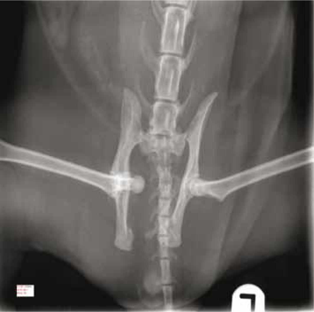
Radiographs revealed a bilateral sacro-iliac luxation with marked cranial displacement of the iliac wings and a right-sided cranio-dorsal hip luxation with marked displacement (Figure 1). Thoracic radiographs were normal.
Twenty-four hours after admission the patient was normothermic, comfortable and his mucous membranes were pale pink; 72 hours after admission his blood glucose had returned to normal. The increase in this parameter was most likely caused by stress-induced hyperglycaemia (Duncan, 1998). On the basis of these findings and after discussion with his owners regarding different treatment options, his owners elected for surgical stabilization of the pelvic fracture.
Pre-surgical patient preparations
The patient was pre-medicated with acepromazine (ACP) 0.01 mg/kg. Meloxicam (Metacam, Boehringer Ingelheim, Berkshire, UK) 0.2 mg/kg subcutaneously and methadone (Comfortan) 0.1 mg/kg intramuscularly were administered to provide analgesia. Once pre-medicated the patient was placed back in his kennel to allow the drug time to reach its peak sedation effect. He was placed on a covered heat source to help decrease the risk of potential hypothermia from lack of movement.
After 30 minutes he was taken into the preparation area of theatre where anaesthesia was induced with propofol (Vetofol, Norbrook, Co. Down, Northern Ireland) and inhalation anaesthesia was maintained after endotracheal intubation using isofluorane (Iso-ba, Schering-plough Animalhealth, Herts, UK). Intravenous fluid therapy was commenced on an infusion pump at a rate of 8 ml/kg/hour using Hartmann's solution (compound sodium lactate, Aqupharm No 11, Animal care Ltd, York, UK).
Intravenous fluid therapy was given to support and balance the circulatory system and prevent it from being compromised by blood loss during the surgery (Daniels, 2010).
In most cases of hindlimb surgery an epidural is routinely administered, however, in this patient an epidural anaesthesia was not undertaken because of the severe bilateral derangement of the overall sacro-iliac and pelvic anatomy.
Both legs were clipped down to each hock and the clipped area was extended to include the perineal region, the proximal tail and the whole circumference of the body caudal to the mid-lumbar region.
All loose fur was vacuumed away. A non-sterile glove and non-sterile bandage were placed on each foot up to the hock to cover the area not clipped. This was done to assist in preventing strike through.
The skin was pre-scrubbed using a solution of Hi-biscrub (chlorhexidine gluconate 4% w/v, Regent Medical Ltd, Manchester, UK) (Box 2).
The patient was then taken to theatre, positioned in lateral recumbency and placed in a thermostatically controlled warm air device (Bair hugger, Advanced Anaesthesia Specialists, Oxfordshire). He was connected to a capnograph and an indirect arterial pressure measurement (oscillometric technique) was used, which was activated to read systolic, diastolic and mean arterial pressure every 3 minutes.
The final stage of surgical skin preparation was then carried out. The patient's left side was prepared for surgery first and once complete he was turned over and the right side was then prepared in the same manner. Prior to draping the patient the surgeon placed a further sterile glove and sterile bandage over each foot to eliminate strike through.
Veterinary surgical procedure
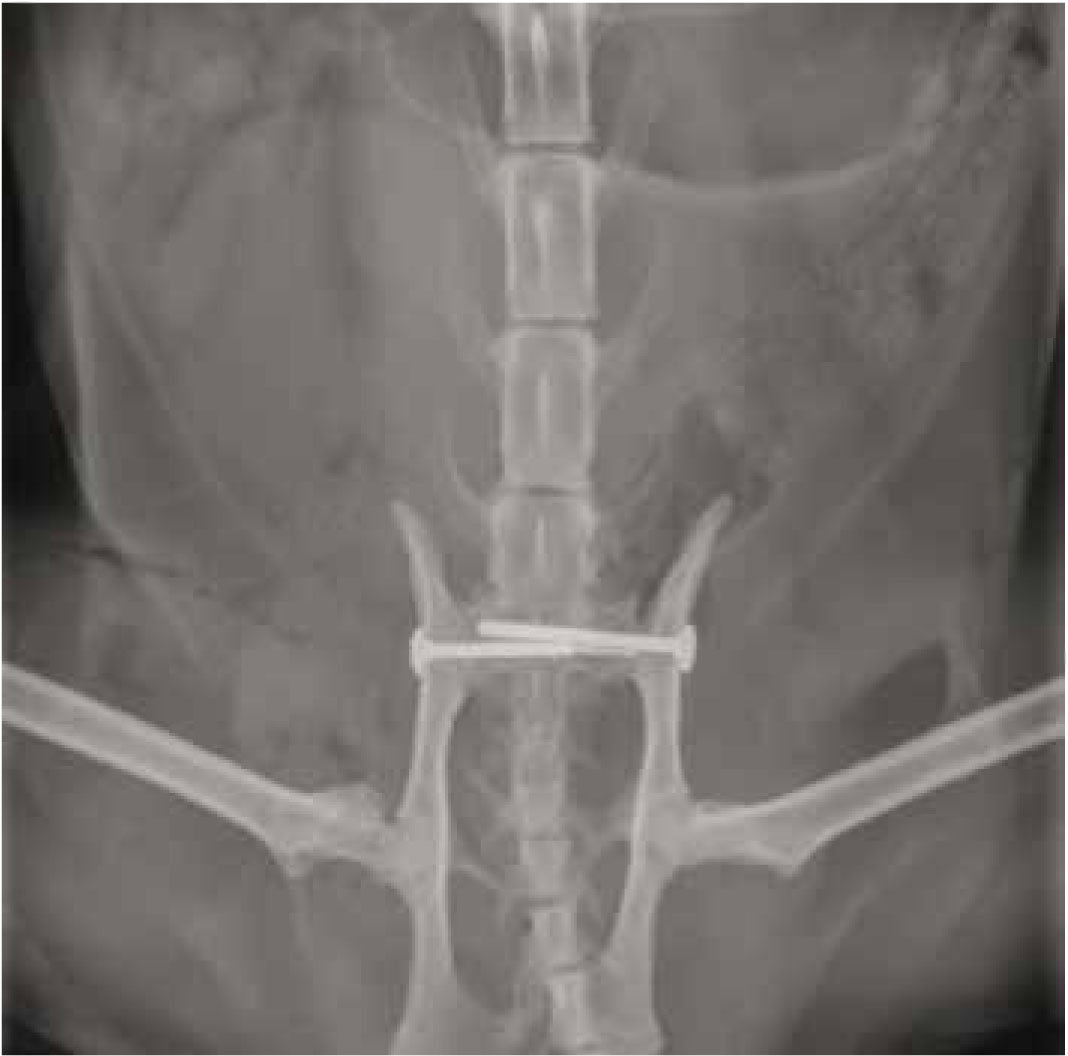
The patient was taken to surgery where the left sacro-iliac joint dislocation was reduced and stabilized using a 2.7 mm lag screw with a plain washer. The same stabilization was undertaken on the right side. The hip joint was reduced and stabilized using a capsulor-rhaphy and using 2-0 polydiaxanone suture (PDS II) (Ethicon, Johnson & Johnson Medical Ltd, Scotland).
Intravenous antibiotics amoxicillin/clavulanate (Augmentin, GlaxoSmithKlein, Middlesex, UK) commenced at a dose of 20 mg/kg and were repeated 2 hours and 4 hours after the initial dose.
Peri-operative antibiotic therapy is routine in extensive orthopaedic procedures, since there is sig-nificant risk of infection around any implants used (Hotson Moore and Garden, 1999).
Post-operative radiography revealed good reduction and implant positioning (Figure 2).
Nursing considerations
On admission to the surgery the patient was immediately treated for shock. This included oxygen therapy, shock rate intravenous fluids for 1 hour and analgesia. He was placed in an incubator which provided him with oxygen and was also thermo controlled to warm him up from his hypothermic state. The use of the incubator allowed him to benefit from oxygen therapy and warmth while keeping him as stress free as possible.
Pain assessment
The patient was showing signs of extreme pain by vocalizing and being aggressive when handled over his pelvic area. A transdermal fentanyl patch (Durogesic, Janssen Pharmaceuticals, Bucks, UK) was placed on the patient's tail base. These patches usually take approximately 12 hours to reach therapeutic efficacy in cats (Kerr, 2007). It is therefore, important to monitor the patient's comfort levels carefully during the period of ef-ficacy, as many animals require additional supplementation with an appropriate opioid that will not compete with the drug administered via the patch (Shales, 2008).
In this case, the patient was given methadone (Comfortan) alongside the patch until the therapeutic levels had been reached. The nurses will spend the most time with the patients and are therefore in an ideal position to comment on the patient's analgesia requirements and response to therapy. Pain is much harder to recognize in cats compared with dogs because cats often do not demonstrate pain signs as overtly as dogs (Orskov, 2010). The author used the Glasgow Composite Measure Pain Scale (Orskov, 2010) as a pain assessment score system to allow a more objective assessment of the patient; this proved beneficial especially when it came to a change of nursing staff.
Training clinical staff to recognize the signs of pain and encouraging the use of pain scales helps to improve everyone's observation skills in this area and will promote a more tailored approach to patient analgesia (Orskov, 2010).
Design of a nursing care plan
A nursing care plan was designed to provide a structural framework of care for all of the nurses dealing with this patient. Planning his care involves ‘nursing to encourage the animal to achieve, maintain or regain maximum independence or to help it adapt to a lower level of independence’ (Joiner, 2000).
Once stable, the patient was placed in a kennel with a thick padded waterproof mattress. This was covered with a urinary incontinence pad to absorb any urine and then covered with a ‘vet bed’ so any urine passed was kept away from the patient. Although he was able to urinate, he was unable to move around his kennel to use a litter tray or move away from any urine passed. It was therefore essential that his bedding was checked regularly to ensure that any urine or faeces was absorbed away from him to minimize urine and faecal scalding.
Although the patient was passing small amounts of urine, it became apparent that he was not totally emptying his bladder; the amount of urine he was passing was small and on palpation after he urinated his bladder was still significant in size (this could potentially result in development of a urinary tract infection). This was most likely because he was unable to stand or squat in a litter tray to pass urine. Over distension of the bladder can result in permanent atony of the detrusor muscle (Petek and Haberer (1995), so his bladder was palpated at least three times daily to ensure the urine passed was not just overflow from an over distended bladder. Manual expression caused him a great deal of distress so he was closely monitored to ensure he was emptying his bladder sufficiently. If this was not the case, a urinary catheter with a closed urinary collection system would have been placed.
Return to function
One of the basic principles of fracture management is an early return to function to minimize fracture disease. This is essential if full limb use following fracture healing is to be achieved (Gemmill, 2007).
Physical therapy has routinely helped human patients recovering from fractures reach their functional goals by helping them regain movement, flex-ibility, strength and balance. Rehabilitation is now commonly provided to small animals recovering from fractures to accomplish similar goals (Doyle, 2004).
Once the fractures have been stabilized the patient is generally more comfortable and should start to use at least one pelvic limb within 24–48 hours after surgery. Post-operative complications are possible so the patient was monitored closely for any changes in his demeanour. His surgical wounds were checked regularly for any signs of infection or dehiscence and both wounds were covered with adhesive dressings to prevent soiling of the area.
The veterinary nurse's role in physiotherapy is to help control inflammation, help control and reduce oedema, manage pain, help maintain and improve range of motion (ROM) and to help strengthen weakened muscles, as well as providing patient contact and bonding (Hill, 2010).
Post-operative physiotherapy commenced the day following surgery. An in-house Association of Chartered Physiotherapists in Animal Therapy (ACPAT) veterinary physiotherapist provided a physiotherapy assessment and treatment plan to be carried out three to four times daily by nursing staff and this was reviewed by the physiotherapist while the patient was in the hospital (6 days) to progress exercises accordingly.
Massage techniques were used to help reduce the swelling in the patient's hind limbs. Passive extension was avoided due to the possibility of hip dislocation.
Facilitated standing (3–4 times daily) using hands for support, on a non-slip surface encouraged him to weight bear through his hindlimbs and this also helped him to regain normal posture and gait. The patient favoured standing on his left hindlimb so swaying exercises were used to encourage him to transfer the weight onto his right hindlimb and then back to his left hindlimb. Facilitated standing and walking are important components of rehabilitation. They both work to improve circulation and lymphatic drainage. The physical act of standing and ambula-tion improves and in some cases retains an animal's mobility, functional capacity and postural balance (Dunning, 2006).
Initially the patient underwent five repetitions of each exercise to avoid fatigue. Repetitions were increased as required depending on the patient's strength gains, as assessed by the physiotherapist.
With increasing demand for post-operative care resulting from more advanced orthopaedic procedures, special emphasis on physical therapeutics can result in a shorter hospital stay and improved patient well-ness (Hill, 2010).
Recommendations for future practice
The purpose of physiotherapy in rehabilitation is to maximize patient recovery, improve patient function and improve the patient's general wellbeing. On busy days it became quite difficult to provide the patient with the regular attention he needed which on some occasions meant he went too long between physiotherapy sessions. The ideal situation would allow for a dedicated physiotherapy nurse to be assigned to these patients, however the financial implication, time constraints and nursing levels within general practice make this an unfeasible option.
Rehabilitation can and should continue at home. Providing owners with education regarding appropriate patient handling and home modifications such as cage confinement allows for a safe return to the home environment. Detailed written instructions for rehabilitation at home promotes owner compliance and accurate completion. This can lead to an increased chance of optimal locomotion of the pelvis and its adjoining anatomy.
The nature of any treatment is influenced by factors such as the extent of the injury, facilities, equipment and the availability of trained personnel.
Financial constraints prevented the patient's owners from leaving him in the hospital for as long as was ideal; ideally he should probably have been kept in a further week to ensure he was having physiotherapy 3–4 times daily, however he was able to stand and walk a short distance and his owners were prepared to continue his physiotherapy at home so he was discharged. His owners were shown the simple techniques and exercises that were being carried out in the hospital on a daily basis. This helped them to appreciate how important certain restrictions (such as cage confine-ment/no jumping/no outdoor mobility) were in order to achieve the best outcome for their pet.
The patient was discharged with the following exercises:
The patient was discharged 6 days after surgery. On discharge he was able to stand independently but was still quite weak on his hindlimbs due to muscle atrophy. With pelvic support, he was able to walk a short distance. The patient returned for regular follow-up appointments and he went on to make a full recovery.
Conclusions
It is essential to consider the entire animal when presented with a fracture case owing to the high incidence of shock, pain and additional soft tissue injuries, all of which should take precedence over fracture assessment in the initial treatment plan. Going into shock means a lack of blood circulating around the body. Basic supportive care for patients in shock includes intravenous fluids, analgesia, external warmth for hypothermia and oxygen therapy and it is necessary to deal with these as a priority. The optimum approach to analgesia requires an ability to recognize and assess pain so that analgesia can be adapted to the individual needs of the patient.
Physiotherapy can help improve flexion and extension of joints, flexibility of muscles, tendons and ligaments and help improve muscle tone and overall limb function. The owners’ active participation in treatment will help to improve the outcome. Unrealistic expectations should be avoided.