
This patient care report highlights the nursing interventions required to manage the jaw fracture patient and critically reflects on alternative care options.
Signalment
Name: Marble
Species: Feline
Breed: Maine Coon cross breed
Sex: Male (neutered)
Age: 5 years
Weight: 6.2 kg
Patient history and assessment
The patient presented to an out of hours emergency centre following a road traffic accident witnessed by the owner. His cardiovascular system was stabilised overnight using intravenous fluid therapy and analgesics and he was referred to the practice's orthopaedic referral centre the following morning for mandibular fracture repair.
On examination, the patient had an unremarkable heart rate of 132 beats per minute and a slightly shallow respiration rate of 40 breaths per minute which constituted tachypnoea. Chest and heart sounds were unremarkable on thoracic auscultation. Marble's mucous membranes were pale pink and moist and his capillary refill time was 1.5 seconds. His temperature was 38.8°c.
Visual examination revealed that Marble had a fractured jaw characterised by lower mandibular deformity (dropped from normal position), blood stained ptyalism (drooling) and evidence of previous epistaxis. There were no other abnormalities detected on orthopaedic and neurological examination.
Jaw fractures
Jaw fractures account for 5% of all fracture cases seen in practice and occur as a result of trauma including road traffic accidents, fighting, falls, kicks and air gun pellets (Glyde and Lidbetter, 2003; Owen et al, 2004). Despite the presence of these obvious and unsightly looking fractures, patients often have more serious concurrent injuries that require stabilisation and investigation prior to surgery. Although not present in this patient, these injuries include upper airway obstruction, central nervous system trauma, pneumothorax and pulmonary contusions which can be life threatening and must take precedence over fracture fixation (Johnson, 2007; Woodbridge and Owen, 2013).
Patient investigations
A 23 g intravenous catheter was placed in the left cephalic vein and blood taken from the jugular vein for an emergency database blood profile. The results showed mild anaemia (27%, reference 31-48%) and a slightly raised alanine transferase (ALT) (145 U/l, reference 33-152 U/l). Marble was pre-medicated using acepromazine 0.05 mg/kg (ACP, Novartis), meloxicam 0.1 mg/kg (Metacam, Boehringer Ingelheim) and methadone 0.3 mg/kg (Physeptone, Martingdale Pharmaceuticals) and anaesthetised with 6 mg/kg propofol (Propoflo, Abbot). Tracheal intubation was performed using a 5 mm un-cuffed red rubber endotracheal tube and a mini-lack circuit attached, and general anaesthesia maintained using 1.4 l/min oxygen and 1.7% isoflurane (Isoflo, AnimalCare LTD). On oral examination there was a central fissure in the hard palate indicating a possible maxillary fracture and a mobile jaw deformity indicating bilateral mandibular fractures. Radiography confirmed bilateral comminuted mandibular fractures. The maxillary fracture was not easily detectable on radiography.
Pre-surgical preparation
The patient was clipped using a size 50 blade on the ventral aspect of the jaw and upper lip regions, extending caudally to the mid neck region and laterally to the level of the eyes. The surgical site was then vacuumed, lint rolled and scrubbed with 50:50 diluted chlorhexidine solution (Hibiscrub, Regent medical). An endotracheal tube was placed via a pharyngostomy incision to divert the airway and improve visualisation of the surgical field. A 12f oesophagostomy feeding tube was also placed before surgery as anorexia or difficulty eating was anticipated for the first few days. The patient received a final scrub in theatre once transferred using the same chlorhexidine solution and a chlorhexidine alcohol solution (Vetasept blue, AnimalCare Ltd) painted on to the skin with gauze swab balls (Zorbs, Millpledge) and forceps.
Surgery
The patient received intravenous fluid therapy throughout the surgery with Hartmann's solution (Aqupharm no.11, AnimalCare LTD) at a rate of 10 ml/kg/hour, reduced to 2 ml/kg/hour during the postoperative recovery period. Although 10 ml/kg/hour rate is still commonly used in practice, there is no real evidence base for using this protocol. The American Animal Hospital Assocation recently published new recommendations for peri-anaesthetic fluid administration suggesting that the administration rate for cats should begin at 2-3 ml/kg/hour and be titrated to the individual requirements of the patient (Davis et al, 2013). Ceforoxime 20 mg/kg (Zinacef, GlaxoSmith, Kline) was administered perioperatively as oral fractures are often contaminated and osteomyelitis a possible complication (Glyde and Lidbetter, 2003; Johnson, 2007).
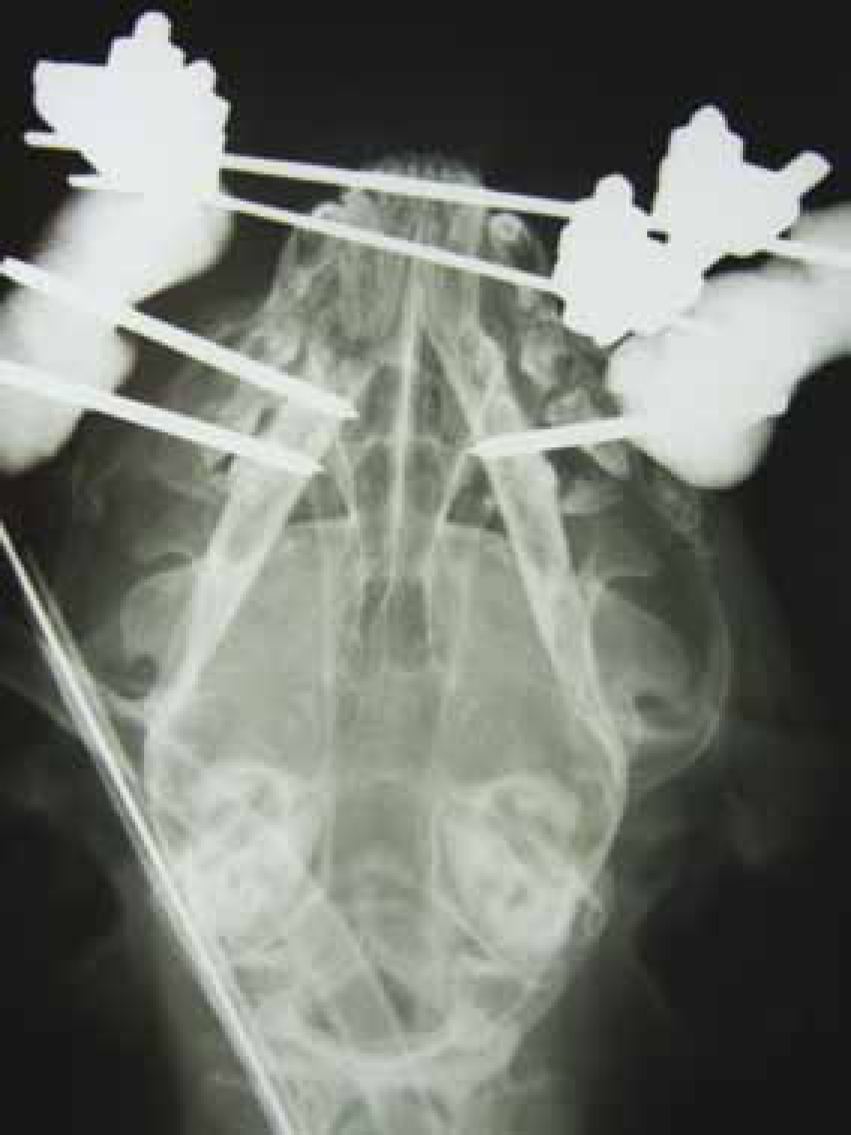
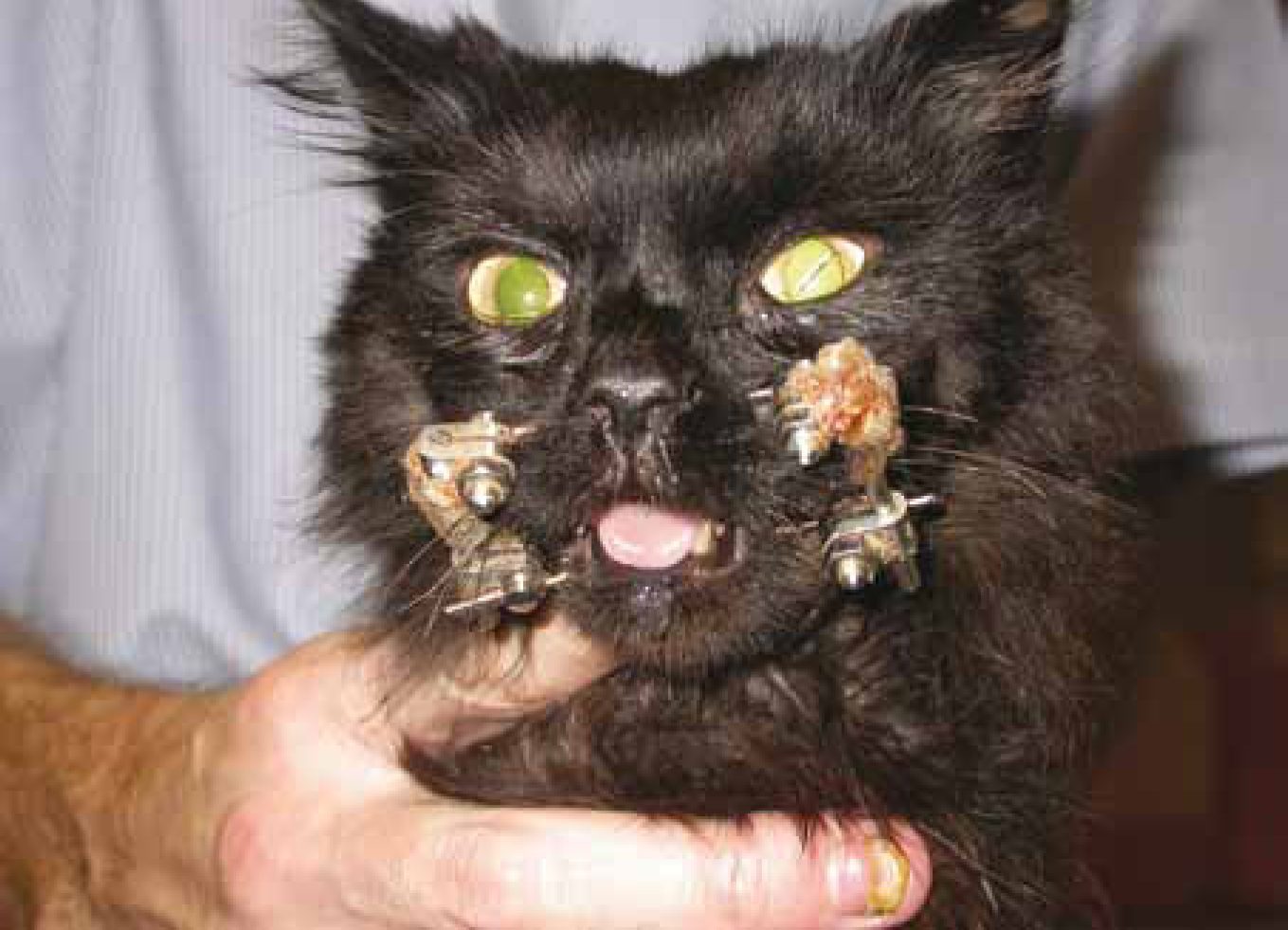
The fractures were stabilised using a mandibulamaxillary external fixator. This involved transverse placement of two positive threaded mid shaft external fixation pins; one through the mandible and one through the maxilla, both caudal to the canine tooth roots (Woodbridge and Owen, 2013) (Figure 1 and 2). A small gap of approximately 5 mm was kept between the opposing incisors to allow the patient to lap and pant as required before the two pins were rigidly fixed together using epoxy putty (Veterinary Instrumentation).
Nursing interventions
The ability to eat, drink and groom is of concern in patients with jaw fractures and these were identified for the purposes of creating Marble's care plan. The care plan was loosely designed around the Orpet and Jeffery Ability Model (2007) (Orpet and Jeffery, 2007) (Table 1) and incorporated into the hospitalisation sheet. Good analgesia was also an important consideration as facial fracture patients often suffer multiple painful injuries.
| NAME: Marble | SPECIES: Feline | BREED: Maine Coon X | ||
|---|---|---|---|---|
| AGE: 5 years | SEX: Male (N) | WEIGHT: 6.2 kg | ||
| REASON FOR ADMISSION: Dog attack: Jaw # (bilateral mandible, mandibular symphysis and maxilla) | ||||
| Ability | Potential problem | Goal | Intervention | Monitoring |
| Eating | Ex fix prevents patient from eating normally, feeding tube in place. Discomfort prevents patient from attempting to eat voluntarily | Ensure patient receives full RER (30 x 5.9 kg)+70 = 247 kcal | Feed patient 300 ml of waltham convalescence diet daily split into 6 meals of 50 ml. Tube should be flushed before and after feed | Weight daily. Tailor calorie intake according to weight changes (+/− 0.5 kg) |
| Drinking | Ex fix prevents patient from drinking normally. Marble usually drinks from pond at home | Ensure patient receives water requirement via tube at feeding times (50 ml/kg/day) | Flush tube before and after feed using water requirement. 300 ml split into 6 tube feeds. 10 ml before, 40 ml after | Monitor urinary output visually. Monitor for signs of dehydration |
| Urination | Marble is an outdoor cat and not used to litter trays | Promote comfortable urination in litter tray | If patient does not like litter or urinates on bed, try soil in litter tray | Monitor urination habits |
| Defaecation | Marble is an outdoor cat and not used to litter trays. Use of opioids and reduced mobility may cause constipation. Concentrated liquid diet may cause diarrhoea | Maintain normal faecal output and consistency | Patient may require micralax enema if constipated | Monitor for signs of constipation or diarrhoea |
| Maintenance of body temperature | Marble does not enjoy being too warm due to thick coat | Maintain normothermia | Ensure kennel area is kept at 18-24°C. Do not provide marble with ‘warm’ bedding such as fleece | Take temperature twice daily |
| Grooming | Ex fix prevents cat from grooming self. Due to thick coat, Marble would typically spend lots of time grooming self. He hates being groomed by someone else! | Ensure face is kept clean. Ensure coat is kept matt free | Clean face using sterile water and cotton buds at least 3 times daily but more frequently if salivating. Groom lightly once daily. Leave marble if he starts to become agitated. Owner is aware we may not be able to groom to the best of our ability | Check coat daily for signs of matting or sores under chin and down ventral neck from salivation |
| Mobilise adequately | Although an outdoor cat, Marble is generally quite lazy and doesn't mind laying in kennel | Ensure Marble remains gently active | Give Marble a small dog kennel due to being large cat. Allow out of kennel in secure room to walk around for 10 minutes once daily | Monitor for signs of boredom |
| Sleep or rest | Pain may prevent adequate rest/sleep. Altough Marble likes dogs, continuous barking may make him stressed | Ensure adequate analgesia and keep stress free | Pain assessment twice daily and more frequently if appears unusually uncomfortable. Keep in separate cat ward if canine patients are too noisy while Marble is in dog cage | Monitor for signs of discomfort |
| Normal behaviour | Marble only enjoys human contact on his terms. He is not an overly affectionate cat | Ensure Marble is kept content despite multiple nursing interventions and interactions required | Keep patient interaction to a minimum unless Marble appears to want human contact | Monitor for signs of stress and agitation during interaction |
Analgesia
A multi-modal, pre-emptive analgesia plan was created for Marble which was initiated with meloxicam and methadone (previously indicated). Methadone was repeated twice more at 4 hour intervals. The patient's comfort level improved (patient became less hunched and quiet and started seeking attention, purring and attempting to groom himself) and methadone was substituted with buprenorphine 0.02 mg/kg (Vetergesic, Alstoe) administered every 6 hours if required for 2 days. Meloxicam was administered once daily for 5 days. The patient was informally pain assessed (visual appraisal of facial expression, body language and vocalisation) every time analgesia was due and 1 hour after drug administration and findings recorded.
Historically, it was believed that animals did not feel pain but veterinary medicine has evolved to have a more accurate understanding of pain and analgesia in animals (Hellyer et al, 2007). The management of pain is considered an important aspect of nursing the surgical patient and nurses are encouraged to actively pain score patients and to treat comfort assessment as the ‘fourth vital sign’ along with temperature, pulse and respiration (Hellyer et al, 2007; Barratt, 2013). In the author's experience the majority of nurses tend to informally pain score patients and at most, write a limited paragraph in the patient's notes of their findings. There have been several pain scoring systems developed in cats to formally identify pain in cats such as the Colorado State University Feline Acute Pain Scale and the Vetergesic Composition Pain Scale for Cats created by Alstoe. A formal means of patient assessment may be considered superior to informal assessment as it provides a structure for nursing hand overs and may be more definitive and less subjective. The use of formal scales encourages nurses to develop awareness and experience in recognition of the clinical signs of pain which include hunched posture, lethargy, anorexia, vocalisation, tail flicking, attacking the surgical site, hypertension, tachycardia and tachypnoea (Barratt, 2013).
Adequate analgesia is not only beneficial for the patient but also to prevent central sensitisation (heightened reactivity to chronic pain) and allodynia (normally non-painful stimuli that cause pain for that patient) which can lead to aversion to non-painful nursing interventions and difficulties in providing complete patient care (Hellyer et al, 2007; Steagall and Monteiro-Steagall, 2013).
Multi-modal analgesia involves the administration of two or more drugs from different classes to provide dynamic pain relief (Steagall and Monteiro-Steagall, 2013). Drugs involved in a multi-modal analgesic protocol may include non steroidal antiinflammatories (NSAIDs), opioids, partial opioids, sedatives and local anaesthetics (Salazar and Leece, 2012). Although not used in this patient, local nerve blocks with peri-neural injection of the maxillary and mental nerves provide local facial analgesia (Salazar and Leece, 2012; Steagall and Monteiro-Steagall, 2013). This technique is underused as it requires accurate dosing and skill (Steagall and Monteiro-Steagall, 2013).
Nutritional management
Oesophageal tube placement is a routine intervention for patients with jaw fracture at the author's practice, especially those with maxillomandibular fixation that are unable to masticate (Johnson, 2007) (Figure 2). The author finds that these patients are initially either completely reluctant to eat, or unable to eat sufficient quantities to meet their resting energy requirement (RER) for several day after surgery (Johnson, 2007; Aldridge and O'Dwyer, 2013). This is likely due to discomfort, pain and decreased ability to smell due to nasal trauma (Glyde and Lidbetter, 2003; Dorricott, 2012). Although many texts recommend initiation of enteral feeding after 3 days of anorexia or 10% bodyweight loss (Chan, 2009; WSAVA Nutritional Assessment Guidelines, 2011; Doricott, 2012; Aldridge and O'Dwyer, 2013), the placing of feeding tubes at the time of surgery pre-empts this need (Firth, 2013). This approach means that tubes can easily be removed if the patient is able to eat post surgery (Glyde and Lidbetter, 2003).
The choice of oesophagostomy tube is popular for jaw fracture patients (Firth, 2013). Nasogastric tubes are often contraindicated in these patients due to facial, nasal and head trauma (Chan, 2009; Hurley and Michel, 2011; Aldridge and O'Dwyer, 2013). If oral feeding by syringe is performed this must be with extreme care as jaw fracture discomfort and reduced ability to orally process food at a normal pace may lead to food aspiration and resultant pneumonia (WSAVA Nutritional Assessment Guidelines, 2011; Dorricott, 2012; Aldridge and O'Dwyer, 2013).
The patient's RER was calculated and a highly digestible liquid diet fed in split meals (see care plan below). Oesophagostomy tube care included daily stoma site cleaning with chlorhexidine, light bandaging to cover the tube, flushing of the tube before and after feeding, and measuring the length of the tube daily to ensure correct tube placement (Orpet and Welsh, 2001a; Aldridge and O'Dwyer, 2013).
The patient was discharged with the feeding tube in place with care instructions for the owner to continue with enteral feeding (Chan, 2009) and she was encouraged to tempt Marble to eat using warmed, aromatic, soft food (Dorricott, 2012). The tube was removed 7 days post surgery when Marble was eating well.
Grooming and facial care
Grooming hospitalised patients though often overlooked when creating a patient care plan, is a very important aspect of patient care as it can provide mental stimulation, promote health and wellbeing, reduce stress and initiate patient-nurse bonding and trust (Orpet and Welsh, 2001b; Aldridge and O'Dwyer, 2013). It was decided that Marble would be checked daily for matted fur and tangles and then groomed as necessary with a metal tooth comb. The principal reason for this approach, rather than full daily grooming was because the patient did not enjoy being groomed and would only tolerate superficial coat brushing before becoming stressed and uncooperative. Some advocate a daily grooming regimen (Orpet and Welsh, 2001b), but the author's experience is that not all animals require or benefit from daily grooming or bathing including some cats, short-coated animals or patients that become easily stressed or scared by unfamiliar people. However, spending time with patients and grooming may help a patient to adapt to its surroundings and gain confidence and trust in nursing staff without medical intervention.
The use of Elizabethan collars is a topic that raises debate among veterinary staff in practice. Some recommend their use in patients like Marble as a prophylactic measure to prevent oesophageal tube and external fixator interference (Orpet and Welsh, 2001b; Aldridge and O'Dwyer, 2013), while others advise the use of collars only as necessary (Owen et al, 2004). The author's experience is that patients may become depressed and frustrated with Elizabethan collars and often tolerate oesophageal feeding tubes very well, especially if they are softly bandaged so that the tube is protected. In this case, Marble was not bothered by his feeding tube or external fixator and did not require an Elizabethan collar.
The patient's face and external fixation bars were cleaned twice daily using diluted 1:1 chlorhexidine solution to reduce risk of infection, and saliva was wiped from his chin as it was noticed or produced. Patients with mandibular fixators, especially man-dibulomaxillary fixators may hypersalivate (Johnson, 2007) which could be considered to contribute to patient depression and frustration due to their fastidiously clean nature. Pin tract discharge and pin loosening is commonly seen in jaw fixation and the risk of osteomyelitis prompts attentive oral care (Owen et al, 2004; Johnson, 2007; Woodbridge and Owen, 2013). Although the practice of daily chlorhexidine mouth rinses is recommended in standard mandibular fixation patients (Johnson, 2007), this was not performed on this patient as the inability to move the jaw reduces the ability to swallow easily and may cause aspiration pneumonia.
Recommendations for future practice
General health and wellbeing can get overlooked in practice, especially when practices are busy and it might be considered more important to administer medication and food than grooming and mental stimulation. The most effective way to ensure that patients receive all aspects of a treatment plan during busy periods is to work as efficiently and productively as possible. Sometimes this may not be possible and the less critical interventions may have to be omitted or delayed. Some care plan activities may also not necessarily require the attention of a qualified nurse. Utilisation of animal care assistants, who have received adequate instruction, is ideal to ensure that components of care plans such as facial care and wiping saliva can be carried out promptly when necessary.
The veterinary team also need to ensure that they prepare thoroughly before anaesthesia for aftercare of jaw fracture patients, as some interventions such as feeding tube placement may be required before surgery is performed.
Conclusion
Jaw fractures alone may not necessarily require the most intensive nursing, however, nurses must be aware that patients with facial fractures often have concurrent injuries that may be more life threatening. The main nursing consideration for these patients is often initiation of early enteral feeding as patients are likely to voluntarily consume less food or not want to eat at all.
The fastidiously clean nature of the feline patient means that catws can become easily depressed and frustrated with drooling and the reduced ability to groom and require intermittent observation and facial care when required.