The skull is unable to expand so any increase in the intracranial pressure (ICP) as a result of an increase in cerebrospinal fluid (CSF) or intracranial blood can have severe consequences such as brain herniation and ultimately death (Bell, 2020). Therefore, there are homeostatic systems in place to tightly regulate the intracranial volume to ensure it remains stable (Bell, 2020). The brain relies on maintenance of intracranial blood volume to meet its high metabolic demands, the cerebral prefusion pressure (CPP) ensures blood is driven into the calvarium to provide oxygen and nutrients and regulates the cerebral blood flow. CPP and mean arterial pressure together determine ICP (Bell, 2020). A trauma to the head will cause an increase in the volume of fluid within the skull because of haemorrhage or oedema; this will cause the CSF and intracranial blood compartments to decrease to maintain ICP. This ultimately results in a reduction in cerebral blood flow and therefore oxygenation and metabolism in the brain (Bell, 2020).
This article will look at the considerations required when anaesthetising the patient following head trauma.
Anaesthetic considerations for patients with head injuries
The veterinary surgeon will decide when anaesthesia is necessary — this may be at the time of injury or may not be until the patient has been stabilised, which may take a few hours or even days. This decision needs to be based on thorough pre-anaesthetic assessments including full clinical examination, clinical history, neurological examination, including the use of the Modified Glasgow Coma Scale (MGCS), and other baseline parameters including haematology, biochemistry, blood glucose, electrolyte status, packed cell volume and urinalysis (McMillan, 2015). The amount of information available will depend on the severity of the head trauma and on whether raised ICP is suspected, because stabilisation may take priority over sample collection. It could even hinder sample collection, as raising a jugular vein for sampling will increase the ICP; this means blood may have to be obtained from peripheral veins instead, which could limit the amount that can be taken, and therefore the number of tests that can be run. Care should be taken when restraining for sampling so as not to inadvertently apply pressure to the neck or cause stress to the patient, which will increase ICP — sedation may be required to do this safely (McMillan, 2015). Priority should be made for samples that will indicate electrolyte imbalances and hydration status, as these should be corrected before anaesthetising the patient (Leece, 2018).
The main aim when anaesthetising patients with raised ICP is to maintain the cerebral blood flow and prevent increases in ICP that could lead to life-threatening brain herniation (Raisis and Musk, 2017). It is imperative that the veterinary nurse or veterinary surgeon anaesthetising the patient understands how cerebral blood flow is regulated and the risk factors and signs of increased ICP, which are summarised below.
When there has been trauma to the head there is an increase in the intracranial volume as a result of haemorrhage and/or cerebral oedema. There is autoregulation within the cranial space which will reduce the production and displace CSF to counteract the increase in volume. This aims to prevent an increase in ICP (Elias et al, 2019). However, once this initial homeostatic mechanism has been exhausted and CSF cannot be displaced further, even a small increase in intracranial volume, such as continued haemorrhage or swelling, will result in large increases in ICP (Vite and Galban, 2018; Bell, 2020).
Normal ICP is 5–12 mmHg, however directly measuring this in practice is challenging, so monitoring for clinical signs of an increase and implementing nursing interventions to prevent increases are recommended.
Compensatory mechanisms are initiated when there is an increase in ICP, however, these will eventually become exhausted and a further increase in intracranial volume will elevate ICP resulting in clinical neurological signs and deterioration of the patient (Plat and Olby, 2017). Signs of raised ICP include: papilloedema, abnormal pulsing of retinal vessels, depression, stupor or coma. Elevated blood pressure with a low heart rate is also a sign of raised ICP and is called the ‘Cushing reflex’. This occurs because of the reduction in cerebral blood flow that occurs to reduce intracranial volume. A reduction in blood flow results in an accumulation of carbon dioxide as a result of reduced perfusion. The rise in carbon dioxide is detected in the brain stem and the sympathetic nervous system responds by increasing mean arterial pressure (MAP), which is then in-turn detected by the baroreceptors resulting in a reflex bradycardia (Bell, 2020).
Anaesthetic concerns
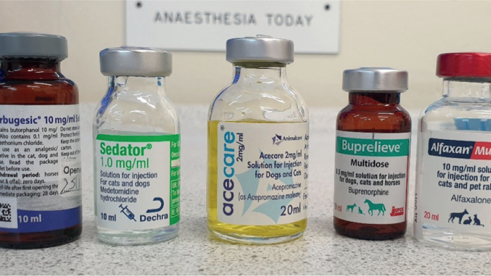
The drugs chosen as either premedication, induction agent or anaesthetic maintenance agent should not increase ICP directly or cause dramatic changes to the patient's blood pressure and should maintain adequate cerebral perfusion (Vite and Galban, 2018). The aims of a premedication are to provide analgesia, reduce stress and anxiety and reduce the dose of induction and maintenance agents required; however, no drug alone will have all these properties, therefore a multimodal approach is required to meet all the patient's needs (Waring, 2017). Figure 1 shows an array of possible sedation and premedication drugs.

Opioids such as methadone and butorphanol do not alter cerebral blood flow or increase ICP. They also have minimal cardiovascular and respiratory depression effects, particularly if used in patients that are in pain and require analgesia (Raisis and Musk, 2017). Potential side effects can also be reduced if the dose is tailored to the individual patient (Raisis and Musk, 2017; Leece, 2018). If the patient is unstable it is recommended by Raisis and Musk (2017) that a short-acting agent that can be titrated to effect is used, such as fentanyl. Care must be taken to avoid morphine or hydromophone as vomiting is undesirable and can cause a rapid increase in ICP (Leece, 2018). Methadone or buprenorphine would be better choices as vomiting is uncommon with these drugs and, unlike mophine, they are licensed for use in cats and dogs. Coughing can similarly cause an increase in ICP, which will be referred to later.
Benzodiazepines, such as diazepam and midazolam, can be useful for reducing anxiety but can cause unpredictable behaviour, excitement and dysphoria (Waring, 2017). They also do not cause any adverse intracranial respiratory or cardiovascular effects (Armitage-Chan et al, 2007). Combining them with an opioid will provide sedation and analgesia in anxious and elderly patients, and they will reduce the overall dose of other agents such as propofol, thus preventing further depression of cardiovascular and respiratory systems (Armitage-Chan et al, 2007). Phenobarbital is also suggested by Raisis and Musk (2017) as a useful premedication in anxious dogs if combined with an opioid such as methadone.
Acepromazine is an anxiolytic and used often as a premedication in healthy animals, however, Raisis and Musk (2017) reported that if intracranial pathology is present acepromazine has been linked to triggering seizure activity. If no intracranial pathology is present then acepromazine itself does not cause or trigger seizure activity (McConnell et al, 2007; Drynan et al, 2012). Acepromazine also causes systemic vasodilation, which may lead to hypotension, and the consequential cerebral vasodilation will lead to increased ICP so should be avoided for this reason alone (Martinez, 2017).
Alpha-2 agonists, such as medetomidine and dexmedetomidine, do not appear to affect ICP in canine or feline patients, however, because they cause marked sedation and significant cardiopulmonary dysfunction they should only be used at very low doses (Armitage-Chan et al, 2007). Leece (2018) suggested medetomidine administered in low doses intramuscularly is preferable over struggling to restrain an aggressive animal for intravenous catheter placement. Low doses are also recommended as alpha-2 agonists induce vomiting in cats at high doses. It is also worth noting they cause increases in MAP and bradycardia, which simulates the Cushing reflex and therefore makes monitoring for raised ICP difficult (Raisis and Musk, 2017).
Ketamine has historically been reported to increase ICP and thus has been avoided in patients with head trauma (Armitage-Chan et al, 2007; Leece, 2018). However, Chang et al (2013) conducted a review of research in human medicine and concluded that the use of ketamine in a controlled ventilation setting, such as under anaesthesia and when used in combination with other sedative drugs, demonstrated no increase in ICP. Unlike other sedatives ketamine is an N-methyl-D-aspartate (NMDA) receptor inhibitor; NMDA receptors are implicated in ischaemic injury so it has been theorised that ketamine may have some beneficial neuroprotective effects (Armitage-Chan et al, 2007). Ketamine also has few cardiovascular or respiratory depressing effects but has been demonstrated to increase cerebral oxygen consumption — these effects can be reduced by co-administration with a gamma aminobutyric acid (GABA) agonist like propofol. In addition, when ketamine is administered with propofol it has even been reported to decrease ICP (Armitage-Chan et al, 2007). Figure 2 is an example of a ketamine constant rate infusion set up.

Induction and endotracheal intubation
Titrating an induction agent to effect, such as alfaxalone or propofol, helps maintain normal ventilation during induction (Warne et al, 2015). The choice of induction agent should be made based on what the veterinary surgeon is most comfortable with using as selecting a drug that they have not used before in an emergency situation may lead to increased errors. Propofol and alfaxalone are the most commonly used induction agents. Propofol maintains cerebral blood flow and decreases ICP by reducing cerebral metabolic rate. The cardiorespiratory effects of propofol are dependent on the dose and rate of administration, meaning that the effects can be lessened by titrating slowly to effect (Raisis and Musk, 2017). Alfaxalone has minimal effect on cerebral blood flow, but also maintains cerebrovascular reactivity to carbon dioxide which is desirable when ICP is increased (Bini et al, 2020).
Co-induction agents are sedatives or analgesics that are given immediately before an induction agent (Leece, 2018). They can be beneficial in these cases as they can help to reduce the dose of the induction agent needed and therefore the associated side effects; this is particularly beneficial in animals that may not be stable enough to be premedicated. Some co-induction agents, such as lidocaine and butorphanol, can also reduce coughing during intubation and other co-induction agents include benzodiazepines and fentanyl (Leece, 2018).
Preoxygenation is vital in head trauma patients to help prevent hypoxaemia as long as it is delivered in a way to minimise stress, however a mask will provide the longest period of time before desaturation compared with oxygen flow-by, and, therefore, a mask should be used if possible (Ambross et al, 2018). It is imperative that an adequate plane of anaesthesia is reached before intubation is attempted in order to prevent provoking a coughing reflex and thus a raise in ICP (Leece, 2018). Lidocaine (1 mg/kg intravenously) can be given to dogs 1 minute before induction and has been shown to reduce ICP as well as help minimise the coughing response (Leece, 2018). Using topical lidocaine before intubating cats, or using a co-induction agent, will also reduce the incidence of coughing, but ensuring an adequate plain of anaesthesia before attempting intubation will be the best way to prevent a cough reflex (Raisis and Musk, 2017).
Maintenance of anaesthesia
Isoflurane causes a slight increase in ICP, more so than sevoflurane, which has been reported to produce no rise in ICP in humans if given at minimum alveolar concentration (MAC) (Rasis and Musk, 2017). Halothane and desflurane should be avoided as both cause marked increases in ICP (Raisis and Musk, 2017). Ideally head trauma patients should be ventilated to normocapnia, which will help reduce the detrimental effects of the inhalation agents (Raisis and Musk, 2017). Total intravenous anaesthesia (TIVA) is suggested by Leece (2018) as the preferred anaesthetic technique over inhalation anaesthesia as it provides good cardiovascular stability. Propofol and alfentanil have been reported to provide good cardiovascular stability and rapid recoveries. Alfaxalone has also been used successfully for craniotomies in canine and feline patients (Leece, 2018).
Monitoring and supportive care
Patients with head trauma and the potential for increased ICP require extensive monitoring (Figure 3), which will ideally include invasive blood pressure monitoring. This will require arterial access, which will require significant skill to place and ongoing intensive care, but will allow continuous direct blood pressure measurement which will be unaffected by hypotension or any cardiac arrythmias (Roberts, 2016). Central venous pressure will be useful to help guide fluid replacement requirements following blood loss, however as discussed previously, there is a risk when placing a central line of increasing ICP because of the compression of the jugular vein needed to insert the catheter (Leece, 2018). An alternative to a central line would be a peripherally placed central line, which can be introduced to the thoracic caudal vena cava via the medial saphenous vein (Leece, 2018).

Intraoperative fluid therapy is essential to maintain normal blood volume and electrolyte balance, and the patient should be normovolaemic before a hyperosmolar solution such as mannitol is administered to reduce ICP (Platt and Olby, 2017). However, excessive fluid therapy will be detrimental as increases in venous pressure may predispose the patient to an increase in ICP (Raisis and Musk, 2017). 0.9% saline is preferable to Hartmann's/lactated ringers solution as these fluids are slightly hypotonic and contain calcium, which has been implicated in secondary brain injury as it facilitates excitotoxin cell damage (Leonard and Kirby, 2002). Prolonged infusion of 0.9% saline, however, may cause hyperchloraemic acidosis (Leece, 2018).
Temperature monitoring is vital to enable maintenance of normal core body temperature. It is considered beneficial to maintain the patient in a hypothermic state in human medicine as it would reduce the cerebral metabolic rate (Raisis and Musk, 2017). However, the adverse effects of hypothermia, including increased risk of infection, increased surgical blood loss, shivering and increased oxygen consumption in recovery, could cause serious complications (Raisis and Musk, 2017). Therefore, this technique should be avoided until there is more veterinary-based evidence for it (Raisis and Musk, 2017). Additionally, hyperthermia should also be avoided as it increases cerebral metabolic rate, which in turn increases cerebral blood flow leading to increased ICP and reductions in CPP (Raisis and Musk, 2017).
Mild head elevation is beneficial to assist venous drainage and prevent increases in ICP. Positioning is therefore important not only in the kennel but also during surgery or imaging, such as magnetic resonance imaging (Figure 4) or computed tomography. The patient should be placed in sternal recumbency, but care should be taken to prevent occlusion of the jugular veins as this will result in a marked increase in ICP (Sturges et al, 2019).

Recovery and postoperative care
A rapid but smooth recovery would be optimal to enable neurological status to be monitored following anaesthesia (Leece, 2018). However, a prolonged recovery may occur in patients with severely increased ICP or in those that have undergone craniotomy as part of their treatment. Ventilation with an air and oxygen mixture would be preferable to 100% oxygen to minimise the risk of oxygen toxicity (Leece, 2018). During the recovery period it is important to minimise excitement and agitation by recovering the patient in a quiet environment that is warm and ideally dimly lit to avoid too much stimulation and increase in blood pressure. Kind and soothing voices and gentle touch are also useful (Raisis and Musk, 2017).
The patient should be warmed as usual in recovery to prevent hypothermia and shivering. Patients may benefit from recovering in sternal recumbency to allow better spontaneous respiratory function; however, sufficient padding will be required to keep the patient comfortable, especially in patients with arthritis. Care should be taken that this padding does not inadvertently put pressure on the jugular veins (Leece, 2018). Extubation should be performed as soon as possible, before the return of the ‘gag’ reflex in dogs, which will reduce the likelihood of a cough (Leece, 2018).
Analgesia is vital during recovery to prevent arterial hypertension that can occur secondary to nociception (Leece, 2018). Although the brain itself does not have any sensory or pain receptors, the skin, periosteum and meninges all have sensory innervation and humans often report headaches post craniotomy, therefore, it would be correct to assume that veterinary patients also experience this (Leece, 2018). A multimodal approach to analgesia would be ideal. Opioids are beneficial, but care needs to be taken to avoid morphine because of the increased risk of vomiting. Opioids will affect pupil size and responsiveness, which needs to be taken into account when performing Modified Glasgow Coma Scale assessment (Leece, 2018)
Careful monitoring of urine output and specific gravity is recommended, especially as head trauma patients are unable to take on oral fluids; therefore, an indwelling urinary catheter may be useful and could be placed under anaesthetic. This will also help with nursing the patient post anaesthesia, especially if they are obtunded and unable to move to pass urine outside (Leece, 2018). There is a high incidence of central diabetes insipidus occurring in humans following head trauma, so it would be reasonable to assume that this can occur in veterinary patients, but may often go undiagnosed (Croton et al, 2019).
Nursing care is paramount post anaesthesia; analgesia such as paracetamol has a good effect on neurological pain but must not be used in cats (Leece, 2018). Lidocaine IV can also be used to provide analgesia as well as reduce ICP. Seizure activity may develop in patients following head trauma and will need to be controlled to prevent further injury. Nutritional support should not be overlooked, particularly in comatose patients and those that may have also sustained facial injury or fractures and that may be unable to eat for themselves (Leece, 2018).
Conclusion
While it will always be the veterinary surgeon that decides the drug protocols for individual cases it is important that veterinary nurses understand the physiology of both the normal and injured brain, including how any drugs administered might affect the patient, and adverse events that the veterinary team can then prepare for. Veterinary nurses are best placed to be responsible for the intensive patient monitoring needed to help in the identification of any signs of deterioration or recovery. Understanding this vital role and being confident to carry it out will make caring for these difficult cases rewarding, and also lead to positive patient outcomes.
KEY POINTS
- The skull is a solid structure that does not allow for expansion in volume of the brain, therefore cerebrospinal fluid (CSF) and blood volume must be regulated to maintain inracranial pressure (ICP). A trauma to the head will affect this homeostatic mechanism with potentially fatal results.
- Anaesthesia following head trauma may be requested by the veterinary surgeon to allow imaging or surgery, however the patient should be thoroughly assessed and stabilised before anaesthesia to reduce potential complications.
- The main aim when anaesthetising patients with head trauma is to maintain cerebral blood flow to ensure the brain has a supply of oxygen and nutrients and carbon dioxide and waste products are removed. Any increases in ICP, which may lead to brain herniation, should also be prevented.
- Choice of pre-anaesthetic drugs and inhalation agents needs to be carefully considered, taking into account potential adverse effects that could cause an increase in ICP. A multimodal approach to a premedication is recommended. Preoxygenation and ensuring an adequate plane of anaesthesia is vital before intubation to prevent the cough reflex which will increase ICP.
- Extensive monitoring is required throughout recovery and hospitalisation. Blood pressure, temperature and mentation should be monitored closely as changes in trends will alert the veterinary nurse to improvements or deteriorations in the patient's condition. Acting swiftly to these changes will give these patients the best chance of survival.