References

Bond R, Riddle A, Mottram L, Beugnet F, Stevenson R. Survey of flea infestation in dogs and cats in the United Kingdom during 2005. Vet Rec. 2007; 160:(15)503-506 https://doi.org/10.1136/vr.160.15.503

Curtis C. Pruritus in dogs and cats part 1: what is pruritus and how do we approach the pruritic patient?. The Veterinary Nurse. 2022; 13:(8)355-360 https://doi.org/10.12968/vetn.2022.13.8.355

Favrot C, Steffan J, Seewald W, Picco F. A prospective study on the clinical features of chronic canine atopic dermatitis and its diagnosis. Vet Dermatol. 2010; 21:(1)23-31 https://doi.org/10.1111/j.1365-3164.2009.00758.x

Franc M, Bouhsira É, Beugnet F. Direct transmission of the cat flea (Ctenocephalides felis) between cats exhibiting social behaviour. Parasite. 2013; 20 https://doi.org/10.1051/parasite/2013050

Halliwell R. Revised nomenclature for veterinary allergy. Vet Immunol Immunopathol. 2006; 114:(3-4)207-208 https://doi.org/10.1016/j.vetimm.2006.08.013

Hill PB, Lo A, Eden CAN Survey of the prevalence, diagnosis and treatment of dermatological conditions in small animals in general practice. Vet Rec. 2006; 158:(16)533-539 https://doi.org/10.1136/vr.158.16.533

Hinkle NC, Koehler PG, Patterson RS. Host grooming efficiency for regulation of cat flea (Siphonaptera: Pulicidae) populations. J Med Entomol. 1998; 35:(3)266-269 https://doi.org/10.1093/jmedent/35.3.266

Miller WH, Griffin CE, Campbell KL. Hypersensitivity disorders, 7th Edition. In: Miller WH, Griffin CE, Campbell KL (eds). Missouri: Elsevier; 2013

Mueller RA. Atopic dermatitis and food-responsive dermatosis, 4th Edition. In: Jackson H, Marsella R (eds). Gloucester, UK: BSAVA Publications; 2021

Park SJ, Ohya F, Yamashita K, Nishifuji K, Iwasaki T. Comparison of response to immunotherapy by intradermal skin test and antigen-specific IgE in canine atopy. J Vet Med Sci. 2000; 62:(9)983-988 https://doi.org/10.1292/jvms.62.983

Rosser EJ Diagnosis of food allergy in dogs. J Am Vet Med Assoc. 1993; 203:(2)259-62

Shaw SE, Kenny MJ, Tasker S, Birtles RJ. Pathogen carriage by the cat flea Ctenocephalides felis (Bouché) in the United Kingdom. Vet Microbiol. 2004; 102:(3-4)183-188 https://doi.org/10.1016/j.vetmic.2004.06.013

Stedman K, Lee K, Hunter S, Rivoire B, McCall C, Wassom D. Measurement of canine IgE using the alpha chain of the human high affinity IgE receptor. Vet Immunol Immunopathol. 2001; 78:(3-4)349-355 https://doi.org/10.1016/S0165-2427(01)00242-2

Pruritus in dogs and cats part 2: allergic causes of pruritus and the allergic patient
02 December 2022
13 mins read
Volume 13 · Issue 10
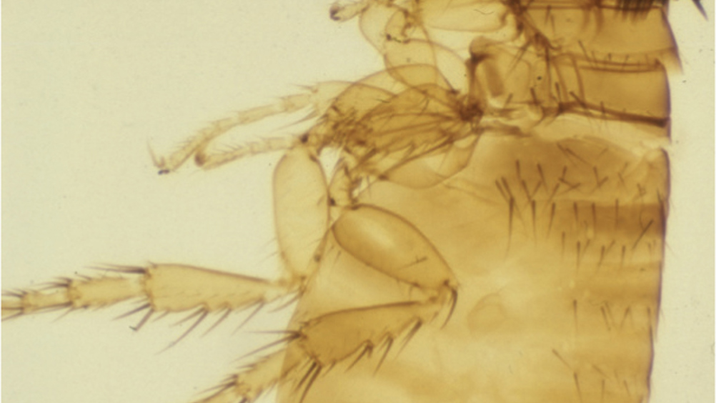
Figure 1. Adult Ctenocephalides felis felis (cat flea).

Abstract

There are many causes of pruritus in domesticated dogs and cats and in this article, the second part of three papers devoted to the subject, the major allergic (hypersensitive) causes are discussed. Despite the tempting tendency to consider ectoparasites a major cause of pruritus in pets, the advent of a number of reliable, safe, effective and long-lasting ectoparasiticides into the veterinary market in recent decades, has meant that the average dog and cat, treated regularly and prophylactically for fleas, ticks and mites, is far less likely to become infested by such parasites. Consequently, allergic causes of itching have become relatively more likely to be seen in general practice. It is therefore important for both veterinary surgeons and nurses to recognise the historical and clinical features of these skin diseases, to allow an appropriate diagnostic and treatment plan to be discussed by the veterinary team and with the client.

In part 1 of this three-part article on pruritus in dogs and cats, pruritus was defined and the general approach to the itchy canine and feline patient was discussed (Curtis, 2022). This second part discusses the allergic causes of pruritus in small animal patients, the approach to these patients, and how a diagnosis of allergic skin disease is made.

The word ‘allergy’ (aka ‘hypersensitivity’) originates from the Greek word ‘allos’, which means ‘other’, pertaining to the pathological process resulting from an aberrant immune response in an allergic individual, following exposure to non-self (‘other’) ‘allergens’. In dogs and cats, as in humans, the commonest allergens that have been described are proteins derived from insects (e.g. flea bite or midge bite hypersensitivity, respectively), the environment (e.g. housedust, dust mites and pollens) and food (e.g. animal or plant proteins) (Miller et al, 2013). These broad groups will be dealt with in turn, and their various historical and clinical manifestations, plus diagnostic and management methods, will be discussed.

Insect (‘flea’) bite hypersensitivity

Pathogenesis and clinical signs

The most frequently observed flea species in both dogs and cats in the UK is the cat flea (Ctenocephalides felis), which was responsible for 93.2% and 98.9% of the fleas found on dogs and cats, respectively, in a 2005 UK survey of over 4000 animals (Bond et al, 2007). This ancient parasite is probably one of the most prolific in the world, having been isolated from over 50 different host species; conversely, individual host species such as the domestic dog can be infested by a variety of flea species (e.g. the dog, hedgehog, rabbit, poultry and even the human flea), demonstrating the truly polyxenous nature of this parasite which undoubtedly contributes to its success.

C. felis is a brown, wingless insect of approximately 3–4 mm in length (Figure 1), with a life cycle comprising oval, larval, pupal and adult stages. Adult C. felis acquire a host and rapidly commence feeding on blood taken directly from capillaries, with females commencing egg-laying within 24–36 hours of this first blood meal and following multiple matings. Once adult fleas have established themselves on a host (within 48 hours), the movement rate to an uninfested host is low (3.7%) (Franc et al, 2013), although host grooming, particularly in cats, appears to be a significant mortality factor, as studies have shown that cats can remove up to 17.6% of their flea burden daily (Hinkle et al, 1998).

Figure 1. Adult Ctenocephalides felis felis (cat flea).

The feeding activity of fleas, during which protein fractions in their saliva are injected into the dermis, can result in moderate to marked pruritus characterised by papules, alopecia and erythema typically affecting the dorsum, rump, caudal ventral abdomen and caudal thighs in hypersensitive canine and feline hosts. Allergic dogs and cats have been shown to develop type I (immediate) and type IV (delayed) hypersensitivity reactions in response to flea saliva, plus late-phase IgE and cutaneous basophil hypersensitivities in the dog (Miller et al, 2013). Affected individuals may develop focal, ulcerated or eroded lesions (aka pyotraumatic dermatitis or ‘hot spots’ in the dog; Figure 2) at the flea feeding sites, or a more generalised, papulocrustous dermatosis, which principally affects the dorsum in cats (aka ‘miliary’ dermatitis) (Figure 3) or the caudo-dorsal rump and caudal thighs in dogs (Figure 4). In cats, diffuse, truncal, symmetrical alopecia may also be observed (Figure 5). Occasionally, lesions of the eosinophilic granuloma complex (EGC) (eosinophilic plaque, linear granuloma and indolent/’rodent’ ulcer) can be linked to flea allergy, although EGC lesions have also been linked to other immunological aberrations. Clinical disease can be seasonal (typically spring through to autumn) or present all year round, depending on the pet's degree of domestication, plus climatic and environmental conditions. In addition to dermatological disease, fleas may transmit other pathogens, for example the tapeworm Dipylidium caninum and, of greater concern, Rickettsia felis, Bartonella henselae and Haemoplasma species, with 17–40% of 121 dogs and cats in one UK study being shown to be polymerase chain reacion (PCR) positive for at least one of these pathogens (Shaw et al, 2004). The zoonotic implications of this finding are obvious, as C. felis has long been known to feed off humans (particularly in the absence of a feline or canine host), and the systemic diseases caused by these organisms are significantly more serious than the more typical pruritic, erythematous wheals most commonly found on the feet, ankles and calves of people inhabiting the environment where a flea-infested pet lives/has lived.

Figure 2. Area of pyotraumatic dermatitis (aka ‘hot spot’) on the rump of a 4-year-old Labrador with flea bite hypersensitivity.
Figure 3. Dorsum of a 2-year-old, Domestic Short Haired cat with miliary dermatitis, alopecia and erythema as signs of flea bite hypersensitivity.
Figure 4. Dorsum and rump of a 5-year-old crossbreed dog showing the typical distribution of pruritus and resultant traumatically-induced alopecia associated with flea bite hypersensitivity.
Figure 5. Dorsum and flanks of a 3-year-old cat with self-traumatically induced alopecia due to flea bite hypersensitivity. Both flanks were similarly affected.

Diagnosis

A review of the client's current and recent flea control methods should be undertaken and it should be borne in mind that all flea life-cycle stages and the insect's faecal material are visible to the human eye, therefore a detailed, direct examination of the haircoat, coat-brushings (either onto white paper or a table top) or the animal's bedding for adult, oval, larval or pupal forms should always be performed as this is often rewarding. If fleas or flea faeces are not observed, intradermal and/or serological testing should be considered, although currently it is difficult to obtain a flea saliva extract for intradermal testing in the UK. In recent years, an enzyme-linked immuno-sorbent assay (ELISA) has been developed which detects circulating levels of antiflea saliva IgE (Stedman et al, 2001), although the author more often prefers to conduct an insecticidal therapeutic trial, which may prove more cost effective (see below)

Treatment

Dogs and cats that are allergic to flea saliva will exhibit signs of pruritus and this should be alleviated with anti-pruritus or anti-inflammatory medications, such as oclacitinib (Apoquel®, Zoetis UK Ltd), lokivetmab (Cytopoint®, Zoetis UK Ltd) or glucocorticoids (e.g. Medrone V®, Zoetis UK Ltd; Prednicare®, Animalcare Ltd). Hopefully, these drugs should only need to be administered short-term, as implementation of, and adherence to, a strict flea control programme will control the problem in the mid-long term. Any flea control programme needs to comprise safe, effective, affordable on/in-animal and environmental insecticides that are easy to administer with long residual activity. The flea's life cycle, and the fact that it is split between host and environment at different stages, needs to be borne in mind when recommending treatment, and clients need to be educated on these points.

Atopy

Pathogenesis and clinical signs

Canine atopic dermatitis is currently defined as a genetically predisposed inflammatory and pruritic allergic skin disease with characteristic clinical features associated with IgE most commonly directed against environmental allergens (Halli-well, 2006). It is a common skin disease of domestic dogs, with an incidence of 5% reported in one UK study (Hill et al, 2006), although other studies have claimed that 10–15% of the global canine population may be affected by this condition. The allergen (which may originate from pollen, dust, dust mites, microbes or dander) is believed to enter the skin directly and be taken up by Langerhans' cells, which roam the epidermis as immune surveillance cells. The offending substance is then presented to T-cells, which become activated and secrete pro-inflammatory and pruritogenic cytokines (e.g. interleukin (IK)-31) and promote the production of IgE antibodies, which coat the surface of mast cells in the dermis. When allergens subsequently cross-link two adjacent Ig E molecules, the mast cell degranulates and a range of pro-inflammatory and vasoactive chemicals are released (e.g. histamine, heparin, leukotrienes and prostaglandins), resulting in redness in the skin (erythema), pruritus and, occasionally oedema/hives (Mueller, 2021).

Typically, dogs and cats are first affected by the disease between 6 months and 3 years of age, but a slightly younger and much older age of onset is occasionally seen. Canine breed dispositions exist in Terriers, English Bulldogs, Boxers, Golden Retrievers and Shar Peis, although no specific feline breeds appear to be predisposed. Clinically, dogs develop reoccurring erythema and pruritus, affecting the face, feet, ears, axillae, abdomen and anus (and vulva in females) either singly, or in any combination as their commonest clinical signs (Figures 69). Secondary infections with bacterial and yeast organisms are frequently observed. In cats, pruritus may be confined to the head and neck (Figure 10) and occasionally otitis is a feature. Feline miliary dermatitis (FMD), a diffuse, papulocrustous lesion, may be present on the dorsum and flanks, and overgrooming of the limbs, dorsum and caudal ventral abdomen is a common feature. Occasionally, lesions of the EGC are also reported.

Figure 6. Left pinna of a 3-year-old atopic, crossbreed dog, showing erythema of its concave surface.
Figure 7. Forefeet of a 2-year-old atopic Miniature Poodle; note the interdigital erythema and the obvious saliva-staining of the fur as a result of excessive licking.
Figure 8. Axillary erythema in a 5-year-old atopic crossbreed dog
Figure 9. Same dog as in Figure 8 — note similar lesions in the inguinal region.
Figure 10. Facial ulceration as a result of excoriation caused by pruritus in an atopic, 4-year-old Domestic Short Haired cat.

Diagnosis

Atopic dermatitis is effectively a diagnosis of exclusion, because there are no specific diagnostic tests for this disease and the major differential diagnoses of flea bite hypersensitivity, cutaneous adverse food reaction, sarcoptic mange, contact irritant/allergic dermatitis, pododemodicosis, trombiculosis, superficial pyoderma and Malassezia dermatitis need to be considered and excluded before a presumed diagnosis of atopy can be made in a patient with suggestive historical and clinical signs (Favrot et al, 2010). Once diagnosed, an attempt to identify the offending allergens using intradermal or serological testing can be made, with the former typically being performed principally by dermatologists. Both tests rely on the detection of either skin-based (i.e. mast-cell bound) or circulating IgE, respectively. Intradermal testing requires the patient to be sedated and an area of fur is clipped from the lateral thoracic wall. Aliquots of 0.05 ml of allergen solution are injected intradermally at sites approximately 1 cm apart, to allow space for a wheal/hive to develop in patients that are reactive to that particular allergen. The test is read at the 15 minute stage, to tie in with the development of a type I hypersensitivity reaction and the size of the wheal is compared with positive (histamine solution) and negative (sterile saline) controls (Figure 11). Serological tests are simpler and require only blood to be harvested and submitted to an appropriate laboratory. Despite several literature reports of poor correlation between the two types of test, immunotherapy success rates based on either test are fairly comparable (Park et al, 2000), although most dermatologists continue to prefer to test the diseased organ in question (i.e. the skin), than to solely rely on a serological antibody test.

Figure 11. Positive skin test result in an atopic dog. Note the large positive reaction in the top left corner (histamine; positive control) and the negative reaction on the right of it where saline was injected as a negative control.

Treatment

Avoidance of the offending environmental allergen(s) is not usually practically possible, although physical removal of allergens, e.g. by regular shampooing of affected areas, can be helpful. A number of different therapeutic options need to be considered to control the pruritus and clinical signs associated with atopic dermatitis and it should also be remembered that atopy is commonly accompanied by secondary cutaneous infections with bacteria (pyoderma) and fungal/yeast organisms (Malassezia dermatitis), which must be considered and controlled with appropriate topical/systemic antimicrobials to improve patients' outcomes.

The author typically grades the patient's pruritus severity as ‘mild’, ‘moderate’ or ‘severe’, and recommends treatment based on this grade. Patients with mild, intermittent or localised disease may respond to topical glucocortoid therapy alone, e.g. hydrocortisone aceponate (Cortavance Spray®, Virbac) or betamethasone (Isaderm Gel®, Dechra Veterinary Products/Betafuse®, Norbrook Laboratories Ltd) on their skin, or triamcinolone (Recicort®, Dechra Veterinary Products) drops in their ears. Such cases may also benefit from oral antihistamines such as chlorpheniramine and, particularly if they have dry skin, from an oral essential fatty acid supplement and moisturising spray. Mild-to-moderately pruritic dogs and cats can benefit from a course of sub-cutaneously injected immunotherapy, based on the results of intradermal and/or serological allergen-specific IgE testing. This is of significant or partial benefit to approximately 40–60% of patients (Miller et al, 2013), but can take several months to take effect, so for welfare reasons, concurrent therapy may be required in its early stages. Moderately affected dogs have the treatment options of oclacitinib (Apoquel®, Zoetis UK Ltd) and lokivetmab (Cytopoint®, Zoetis UK Ltd), which interfere with the production of, and the binding of IL-31 (see earlier), respectively, and both cats and dogs can be given cyclosporine (e.g. Atopica®, Elanco UK AH Ltd), which affects T-lymphocyte function. Patients with severe disease often need to be treated with cyclosporine or systemic glucocorticoids, either by the oral or the injectable route. Given that treatment for atopic dermatitis is usually a lifelong requirement, the side-effects and costs of any drug used to control it need to be borne in mind and discussed with the client and one should always aim to use the minimal dosages necessary for satisfactory control.

Cutaneous adverse food reactions

Pathogenesis and clinical signs

Compared with the wealth of information available on, and research effort devoted to, the subjects of both flea and environmental allergen hypersensitivity, relatively little is known about the true pathogenesis of cutaneous adverse food reactions (CAFRs). A reliable, reproducible true allergic pathomechanism for this condition has not been established, and consequently veterinary dermatologists are cautious about the terminology used to describe food-responsive skin diseases, preferring terms like CAFR or food ‘intolerance’, to food ‘allergy’. Dogs and cats that have skin disease as a manifestation of an adverse reaction to food display clinical signs that are virtually identical to those previously described for atopic dermatitis, and the reader is referred back to these. Occasionally, an extra clue in the history exists in patients that may have a tendency for bouts of intermittent diarrhoea or flatulence, but this is not always the case.

Diagnosis

Serological and intradermal testing for the detection of food-specific antibodies are not reliable in the diagnosis of CAFR and are not recommended (Mueller, 2021). The ‘gold-standard’ test is the novel, limited ingredient, elimination diet and this needs to be compiled following detailed questioning of the owner on the foodstuffs that their pet is fed on, plus treats, left-overs and supplements (which may contain for example gelatin in the capsule) etc. Whether the pet is offered milk to drink is important to establish and also, particularly in cats, whether they hunt and/or potentially visit neighbours' houses, where they may have access to other sources of food. For the latter reasons, it is often recommended that cats are confined indoors during the diet trial period, although this may have welfare implications for some animals. Typically, proteins of animal origin are the most likely foodstuffs to trigger CAFRs, although wheat, soy and rice sensitivities have been identified in dogs (Miller et al, 2013). The novel home-cooked, or commercial diet to be offered should therefore be based on a protein source that the patient has had no prior exposure to, or a vegetarian diet is an option for dogs. Clients should be asked to provide full details of the ingredient lists of all varieties of foods and treats that they offer to their pets, to enable the clinician to compile a restricted, single, novel protein and single carbohydrate diet that should be fed exclusively for at least 3–6 weeks, although some studies claim that a small percentage of dogs with CAFR may be misdiagnosed if the trial is not continued for a total of 10 weeks (Rosser, 1993). Water should be the only drink offered throughout the trial. If the patient's skin disease improves, then the second, or ‘re-challenge’ phase of the trial needs to be undertaken, during which the client re-offers their pet all of their original foods (of all varieties and flavours), plus their usual treats and leftovers etc, plus milk to drink, if they used to have it. This phase should last approximately 1–2 weeks and if the animal becomes pruritic again during that period, the third and final ‘rescue’ phase should be implemented, in which the client again offers only the restricted diet and water, and hopefully reproduces the improvement in the pet's condition. In the UK there is a temperate climate with seasonal variations in pollen counts and insect exposure, in addition clinicians may need to prescribe antimicrobials and even short-term anti-inflammatory/anti-pruritus medications to some patients at the start of a diet trial, so is imperative that the ‘re-challenge’ and ‘rescue’ phases of the trial are undertaken, otherwise variations in the level of the patient's pruritus may be attributable to these other factors and may be inappropriately credited to the change in diet, resulting in misdiagnosis of CAFR.

Treatment

Once a diagnosis of CAFR is made, most clients are happy to leave their pet on the hypoallergenic diet that they have identified, in the hope that they will remain comfortable and that their pruritus will continue to be controlled. If a home-cooked diet has been used during the trial, they may wish to switch to an ingredient-matched commercial diet, for convenience, but if they prefer to continue cooking for their pet, they can seek advice about a suitable vitamin and mineral supplement that may need to be offered, as home-prepared single animal protein and carbohydrate diets may be deficient in some nutrients. Some clients may want to attempt to identify the offending foodstuff, and they can do so by feeding the restriction diet and adding in a single protein source for a couple of weeks at a time (e.g. cooked chicken, then beef, then fish etc.), and keeping a food ‘diary’, but more often they seem content to just continue to offer their pet the hypoallergenic diet that they have responded to, and to sequentially offer a few different treats in a similar way, so that the animal has a small selection of ‘safe’ food items that can be used as rewards.

Conclusion

In modern general practice, because of regular use of efficient ectoparasiticides, the main cause of ectoparasite-linked pruritus likely to be encountered is a hypersensitivity to flea saliva, as opposed to a primary, major infestation with fleas or other parasites that affect the skin. The incidence of allergic skin disease in people and animals is believed to be increasing, and particularly with respect to environmental and dietary ‘allergens’; hence the clinical signs of these diseases must be borne in mind when presented with a pruritic dog or cat. It is also important to remember that serological and/or intradermal allergy tests form only part of the diagnostic approach to allergic skin disease, and should be reserved for patients whose historical and clinical features are consistent with a hypersensitivity disorder and in whom all other potential causes of pruritus have already been excluded.

In the third and final part of this series of articles on canine and feline pruritus, the ectoparasitic and infectious/microbial causes of this clinical sign will be discussed and the various ways of managing these conditions will be addressed.

KEY POINTS

  • Allergic skin disease is, by definition, pruritic, so clients' funds should not be wasted performing tests that are indicated for allergies, or by supplying anti-itch medications.
  • Very small amounts of allergen (e.g. flea saliva) are needed to trigger and maintain an allergic reaction, so any attempts at avoidance of the offending allergen as a means of controlling clinical signs, must be absolute and thorough. This is achievable with flea and dietary allergens, but is rarely possible with environmental allergens, e.g. dust, dust mite or pollens.
  • When compiling a flea control programme, remember to include all in-contact dogs, cats and other mammals (e.g. rabbits), as fleas are not host-specific and appropriate, species-licensed insecticides for co-habiting pets should be recommended, in addition to treatment of the environment.
  • Diet trials need to be continued for a few weeks beyond the use of any other concurrent therapy the patient is receiving (e.g. antimicrobials or anti-inflammatories), to ensure that it is not simply the concurrent medication that is responsible for the improvement. The re-challenge and second rescue phases of the diet trial are mandatory steps in making a definitive diagnosis.
  • Currently, in veterinary medicine, allergies can only ever be ‘managed’, as opposed to ‘cured’, as the tendency for the patient to develop a reaction to the offending allergen(s) is likely to be lifelong. Clients should be made aware of this fact and their expectations managed, as a major reason for them becoming frustrated with their persistently itchy pet, is a misunderstanding of the fact that their disease is chronic in nature. It is important, therefore, to prepare them for regular visits to the veterinary practice and make them aware of the fees associated with keeping their pet comfortable.