How to examine, take samples and prepare slides for diagnosis of canine ear disorders

Abstract
Veterinary nurses play a vital role in ear examination, supporting veterinary surgeons in making their diagnosis. This article provides an outline on how to examine the ears of a canine patient, how to take a swab for cytology and how to prepare slides for examination.
Otitis externa and skin disorders in dogs are named as some of the most common issues presented to small animal, first opinion veterinary practices in the UK (O'Neill et al. 2021). Paterson (2020) states that ear cytology is one of the most important steps in investigating otitis externa. For this reason, it is important that nurses are confident in the process of examining ears, taking samples for diagnostic testing, and preparing slides for examination under the microscope. This guide assumes that a thorough history has already been taken from the owner and that a full health check has been carried out of the rest of the patient's body.
It is important to have all the equipment to hand before beginning the examination. This includes:
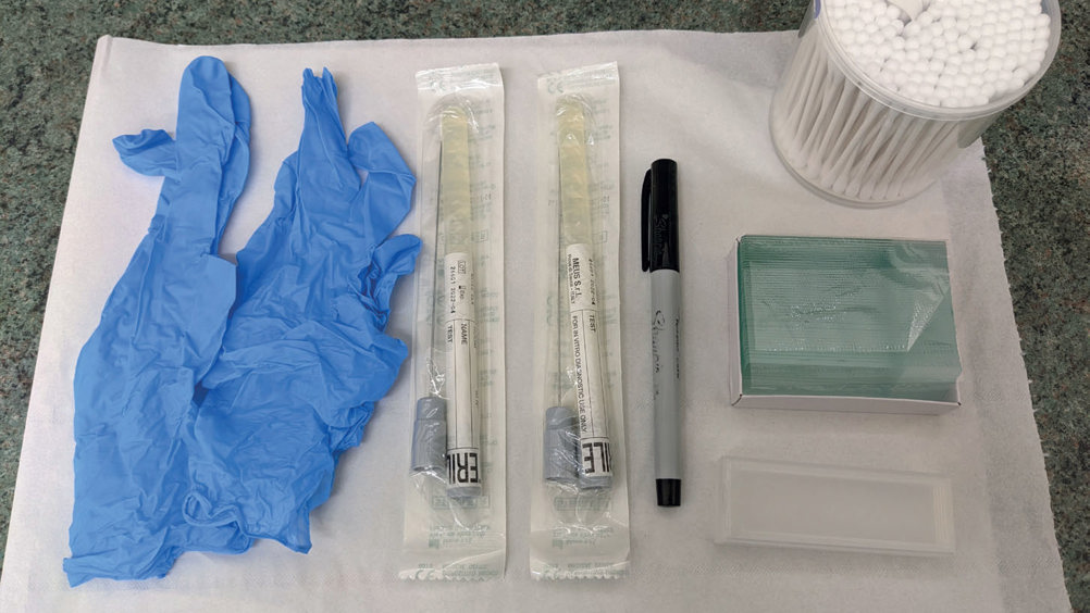
Figure 1 shows the equipment that should be prepared before the examination and sampling begins. Ensure all equipment is in good working order (e.g. the bulb of otoscope is working), and that the equipment is clean. Specula must be cleaned and disinfected thoroughly between patients, and you should use a separate, clean speculum (these can and should be disinfected between patients) for each ear to prevent cross contamination.
Register now to continue reading
Thank you for visiting The Veterinary Nurse and reading some of our peer-reviewed content for veterinary professionals. To continue reading this article, please register today.

