References

Balsa IM, Culp WT Wound care. Vet Clin North Am Small Anim Pract. 2015; 45:(5)1049-1065 https://doi.org/10.1016/j.cvsm.2015.04.009

Bloor C Pain management in critically ill patients. Vet Nurse. 2012; 3:(8)469-530 https://doi.org/10.12968/vetn.2012.3.8.494

Demetriou J, Stein S Causes and management of complications in wound healing. In Pract. 2011; 33:(8)392-400 https://doi.org/10.1136/inp.d5353

Hollis G Good foundations – the structure of wound healing. Equine Health. 2014; 17:20-24 https://doi.org/10.12968/eqhe.2014.1.17.20

Hosgood G Chapter 1: The biology of wound healing, 2nd edn. In: Williams J, Moores A (eds). Gloucester: BSAVA; 2009

Basic principles of wound care. Today's Veterinary Nurse. https://todaysveterinarynurse.com/wound-management/basic-principles-of-wound-care/ (accessed 6 September 2024)

Approach to the bleeding patient. 2011. https://www.vin.com/apputil/content/defaultadv1.aspx?id=5124325&pid=11343&print=1 (accessed 6 September 2024)

LaPelusa A, Dave HD Physiology, hemostasis.StatPearls. Treasure Island (FL): StatPearls Publishing; 2024

Mickelson MA, Mans C, Colopy SA Principles of wound management and wound healing in exotic pets. Vet Clin North Am Exot Anim Pract. 2016; 19:(1)33-53 https://doi.org/10.1016/j.cvex.2015.08.002

Starybrat D, Fenner J, Tappin S Bleeding disorders: causes and treatments. Vet Irel J. 2016; 6:(9)504-521

Waldron DR, Zimmerman-Pope N Chapter 21: Superficial skin wounds, 3rd ed. In: Slatter D (ed). Philadelphia(PA): Elsevier Saunders; 2003

Reconstructive surgery – considerations and planning. https://www.vin.com/doc/?id=5709887&meta=generic (accessed 25 November 2024)

Yon E How to reduce the risk of surgical site infections. Vet Nurse. 2019; 10:(1)42-46 https://doi.org/10.12968/vetn.2019.10.1.42

Wound healing for small animal practitioners: a refresher – part one
02 December 2024
11 mins read
Volume 15 · Issue 10

Abstract

Understanding normal anatomy and physiology is essential for veterinary nurses to recognise the pathophysiology of injury and disease. Wound healing is a physiological process that veterinary nurses frequently observe and manage in practice. This article reviews the wound healing process and highlights its clinical relevance throughout. It will be followed by two subsequent articles addressing wound management protocols in dogs, cats and exotic pet species.

In daily practice, whether in first-opinion or specialist referral settings, veterinary nurses encounter patients with wounds almost every day. These may range from uncomplicated surgical wounds to more complex traumatic wounds. A fundamental understanding of wound classification, the physiology of wound healing, and the principles of wound management, is essential for veterinary nurses to deliver high standards of patient care. This article provides an overview of wound healing in commonly encountered species in small animal practice, with useful comparisons between species. It is the first in a three-part series, which will also explore current wound management protocols for small animals and exotic pet species.

Firstly, it is important to define what is meant by a ‘wound.’ A wound is a disruption of the normal continuity of a body structure (Waldron and Zimmerman-Pope, 2003). While a wound is often thought of as a break in the skin surface—true for open wounds—wounds can also be closed, such as haematomas or contusions. Wounds may be superficial, affecting only the top skin layers (eg, a minor abrasion), or deep, penetrating the skin and potentially involving structures like muscle and bone, as seen in deep puncture wounds or degloving injuries.

Understanding why wounds pose a risk to a patient's health requires consideration of the skin's role. In most animals, the skin is the largest organ in the body and performs numerous functions, the most critical being its role as a barrier between the external environment and the internal body. This barrier is vital for preventing harmful substances and pathogens from entering the body. When the skin is compromised, a direct pathway is created for these substances and pathogens, greatly increasing the risk of infection and disease.

The skin itself is composed of three layers; the epidermis, dermis and hypodermis (also known as the subcutis/subcutaneous layer). For the consideration of wound healing, it is important to consider vascular supply as this is what will enable supply of important substances and nutrients to the wound bed. Dogs and cats have direct cutaneous arteries originating from deeper tissue and penetrate through to the skin; small branches of these arteries supply feed to the dermis (Walker, 2013).

Normal wound healing process: cats and dogs

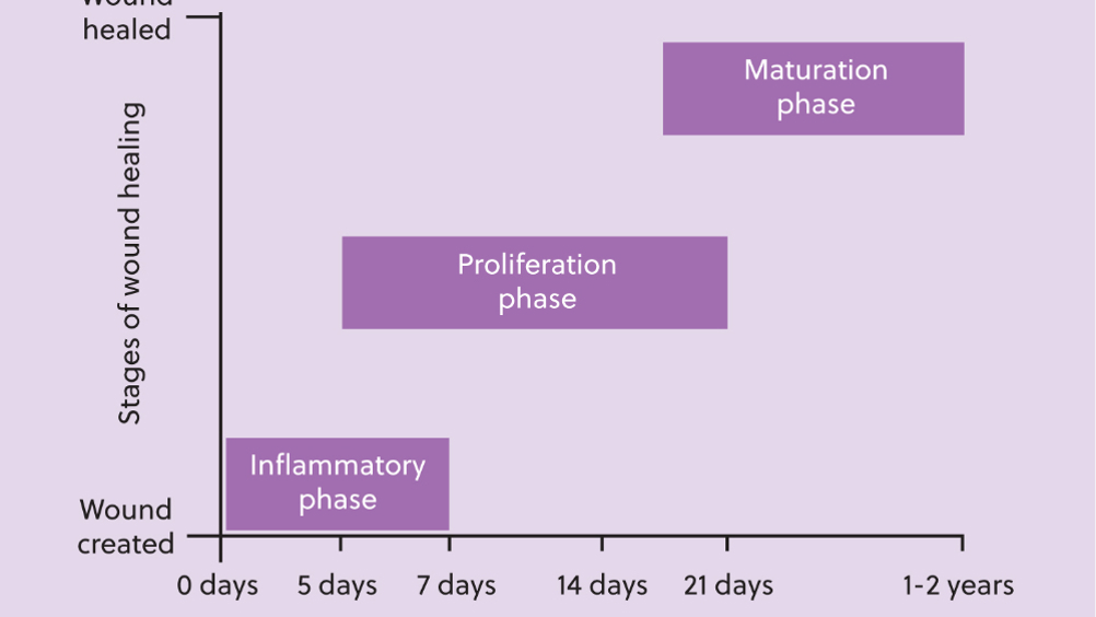
The wound healing process involves four key phases: bleeding, inflammatory, proliferation, and the maturation/remodelling phases (key events are outlined in Table 1). These phases are often assumed to be linear, with one phase clearly following the next, but this is not always the case; phases often overlap with one phase beginning before the previous phase has finished (Hosgood, 2009) (Figure 1).


Table 1. Key events during phases of wound healing
Healing phase Key events
Haemostasis
  • Immediate vasoconstriction proceeded by vasodilation
  • Formation of fibrin clot
Inflammatory
  • Migration of neutrophils, macrophages, fibroblasts and endothelial cells.
  • Signs of inflammation
Granulation
  • Early repair commences (approximately 3-5 days) after wound created
  • Granulation tissue formation and epithelialisation
  • Wound contraction
Maturation
  • Starts approximately 15 days after wound created and may last for up to 12 months
  • Collagen deposited
  • Scar tissue forms
  • 80% of wound strength achieved after 3 months
(Adapted from: Demetriou and Stein, 2011)
Figure 1. Timeline of wound healing.

Veterinary nurses should understand the physiology of these steps to allow them to be able to effectively manage wounds at different stages of the wound healing process.

Haemostasis

Haemostasis refers to the cessation of bleeding (LaPelusa and Dave, 2023). When a wound occurs, blood vessel damage typically results in bleeding. This damage triggers primary haemostasis, a process comprising three steps:

  • Vasoconstriction: the damaged blood vessels constrict to reduce blood loss.
  • Platelet aggregation: platelets aggregate at the site of damage to form a temporary platelet plug. This plug serves as a provisional barrier and provides a foundation for the development of a blood clot.
  • Coagulation cascade: the coagulation cascade is activated, leading to the formation of a fibrin plug (blood clot).

The practical implications of haemostasis are important to consider. Platelets are critical to the process, so a patient with thrombocytopaenia will likely struggle to control bleeding because of an insufficient number of platelets. Without enough platelets, a stable platelet plug cannot form (Starybrat et al, 2016). Additionally, fibrinogen and von Willebrand factor play essential roles in sticking platelets together to facilitate clot formation. Patients with von Willebrand's disease, for instance, may have difficulty forming a platelet plug and are therefore prone to prolonged bleeding (Starybrat et al, 2016).

In cases where a patient cannot control bleeding and haemorrhaging occurs, intervention may be necessary. Methods of achieving local haemostasis include applying wound pressure, ligation, or using topical agents (Kohn, 2011).

When blood vessels are damaged, local cells and tissues become starved and devitalised (Hollis, 2014). For this reason, achieving haemostasis quickly is critical to allow the healing process to progress.

In the initial stages of wound healing, an extracellular matrix (ECM) forms, facilitating the entry of key cells such as neutrophils, macrophages, endothelial cells and fibroblasts into the wound (Hosgood, 2009). The ECM plays essential roles in reducing blood loss, acting as a barrier to infection and fluid loss, and providing a basic scaffold for the early organisation of wound healing (Hosgood, 2009).

Inflammatory/debridement phase

The key cells involved in this phase are neutrophils and macrophages. These cells essentially perform a ‘clean up’ operation, clearing away dead cells, bacteria and protein from the wound bed. Both macrophages and neutrophils ‘ingest’ unhelpful substances, such as those just listed by phagocytosis, in addition to releasing pro-inflammatory cytokines which signal to the immune system to initiate an inflammatory response (Hosgood, 2009). This results in slough and exudate production. Although exudation is often associated with surgical site infection (Yon, 2019), it is an expected and normal part of the healing process and is evidence of the inflammatory phase occurring (Hollis, 2014) (Figure 2). Prolonged periods of exudation, foul smelling or yellow/green exudation is a cause for concern and should be monitored.

Figure 2. Clear exudate can be observed in this wound, with evidence of damaged and necrotic tissue in the cranial wound.

Proliferation/granulation/repair phase

The key activities here are angiogenesis (formation of new blood vessels) and the formation of granulation tissue (Jager, 2020). Granulation tissue, the red-pink tissue that fills the wound bed (Figure 3), is comprised of new capillaries, fibroblasts and fibrous connective tissue (Hosgood, 2009). Fibroblasts produce elastin and collagen, essential for elasticity and wound strength (Balsa and Culp, 2015). The role of granulation tissue is outlined in Figure 4. The presence of granulation tissue is a positive indicator for the health of the wound bed, suggesting adequate blood supply and nutrient delivery. In practice, close monitoring of the wound bed to identify the presence of granulation tissue is essential for determining the progress of the wound healing process, which will impact decision making about ongoing management.

Figure 3. Red-pink granulation tissue fills the wound bed.
Figure 4. Wound moving from proliferative to maturation phase of wound healing.

During the granulation phase, wound contraction also starts to occur (Jager, 2020); the wound size starts to decrease. As this happens, the patient may experience more pain and an increase in attempts to interfere with the wound might be observed. Ensuring adequate analgesia and prevention of interference with appropriate dressings and a buster collar are key. In the author's experience, medical tshirts are less effective at preventing interference because of patients licking and chewing at the material, causing damage or soaking the fabric with saliva. Once the fabric is wet, there is a direct route for bacteria to contaminate the wound. The same applies with dressings, hence the suggestion for a two-pronged approach with dressings and buster collars.

Epithelialisation is the formation of new skin cells. These start to proliferate at the wound edges and will then begin to cover the wound. Hosgood (2009) outlines that in clean, incised wounds, epithelialisation can be completed within as little as 24-48 hours of the wound being created. In larger, full thickness wounds, the process does not begin before approximately 4-5 days after the wound is created (Hosgood, 2009). Again, these timelines highlight the importance of wound monitoring; visible epithelialisation is an indicator of wound health. Depending upon the method of closure, observation of new epidermis may influence the point at which a wound is surgically closed.

Remodelling/maturation phase

In this phase, granulation tissue matures and the number of cells it contains reduces; fibroblasts and endothelial cells begin to die (Hosgood, 2009). The collagen content of the wound begins to decrease, although collagen fibre bundles become thicker, improving the strength of the scar tissue (Hosgood, 2009). A scar has a maximum of 70-80% strength of normal tissue (Hosgood, 2009; Jager, 2020). Figure 4 demonstrates a wound progressing from the proliferation to maturation phase of wound healing. Although the central area of the wound is proliferating, scar tissue is evident at the edges of the wound and the wound has significantly reduced in size. Figure 5 demonstrates a wound in the maturation phase; it is completely closed with scar tissue evident.

Figure 5. All open areas of the wound have now closed and a scar is observed. This wound is now in the maturation phase of healing.

Roles of granulation tissue (Adapted from: Hosgood, 2009)

  • Fills the tissue deficit of the wound
  • Offers protection to the wound bed
  • Provides a physical barrier to infection
  • Provides a framework for epithelialisation
  • Containes myfibroblasts that are important for wound contraction.

The length of time for wound healing is linked with presence of factors affecting wound healing such as size and depth of the wound, level of contamination, closure technique and effectiveness of management; these will be discussed below.

Total capacity for wound healing does differ between cats and dogs; all of the above apply to both species, however, cats have slightly different vasculature and the production of granulation tissue, epithelialisation and total healing are reduced in cats (Demetriou and Stein, 2011).

Comparative wound healing physiology

In 2024, an estimated 1.5 million indoor birds, 1.3 million domestic fowl, 700 000 tortoises and turtles, and 600 000 snakes were kept as pets in the UK (Food, 2024). Consequently, a significant portion of these animals will likely require veterinary care at some stage.

Understanding how the skin of reptiles and birds differs from that of mammals provides valuable insight into appropriate wound treatment, closure methods, and bandaging options. With over 10 000 species of birds and reptiles, there are notable variations; however, this discussion will focus on key anatomical differences in commonly seen pet species.

Birds

Bird skin has little to no hypodermis layer; it is incredibly thin. As a result of this, it is loosely attached in places to the underneath structures. Places such as the lower leg however attach directly to bone. The skin has an outer epidermal layer composed of three main layers from inside to out: the germinatory layer; the maturation layer and the cornified layer. Their dermis layer, compared to mammals, is also reduced. As a result, birds have a significantly reduced elasticity to the skin. Their skin also lacks sweat glands (Girling, 2013).

The propatagium is a triangular fold of feathered skin on the leading edge of the wing, between the shoulder and carpal joints. In its cranial-free edge is the elastic propatagial ligament. These structures maintain the leading edge of the aerofoil structure of the wing and their integrity is essential for flight (Jones and Dodd, 2012). Birds commonly sustain skin injuries in this area and thus it is important to be aware of this anatomy to ensure promotion of wound healing and effective wound management.

A distinct feature that is unique to birds, is of course, their feathers. They develop from feather follicles in the epithelial cells, like hair in mammals and scales in reptiles, and are made of the same material: keratin (Aspinall et al, 2015). For surgical preparations or wound closure, plucking feathers in the area for asepsis is important, similar to shaving in mammals. However, it is important to note that this procedure can be very painful, so sedation or anesthesia, along with pain relief, must be provided.

During the formation of a new feather, a shaft is created which fills with blood; this is called a blood feather. If damaged, this can cause a significant amount of bleeding. Depending on the bird's size this volume of blood loss can be life threatening. Care must be taken when handling birds with blood feathers (Figure 6).

Figure 6. Blood feather in a bird.

Some areas of the skin are devoid of feathers; these are normal and called apteria, most notably over the right jugular in many species of bird, including parrots. This does not occur in all species; waterfowl and pigeons for example do not have an apteria, but parrots and corvids do.

Reptiles

Reptiles have scales that form from the folding of the outer epidermal layer. Size of scales and textures varies. Some species have scales with ridges on their surface to add greater grip, other species have smooth scales. The elastic property of skin varies between species, some having very little, some having a lot. The reptile skin has little to no skin glands. The outer layer, known as the stratum corneum, is formed of three layers of dead cells filled with keratin (Girling, 2014).

Snakes and lizards undergo shedding, and once a wound has healed and scar tissue has formed, it may affect the animal's ability to shed. For example, snakes should shed completely, but if scared, they may need assistance as the skin may not come off easily. Part three will cover this in more detail.

Chelonia

Tortoises and turtles have a shell, making them unique to all other animals. There is a common misconception regarding the anatomy of the shell that they do not feel pain when it is injured, equating it to hair and nails of other creatures as it is made up is of the same substance, keratin. This is not the case; the shell is made of fused living dermal bone covered by keratinised epidermis. The top of the shell (carapace) is where thoracic, lumber and sacral vertebrae and ribs are flattened and fused.

The carapace and bottom of the shell (plastron) are attached together between the fore and hind limbs by the pillars of the shell. The individual sections of the shell are called scutes and are made up of shell epidermis (Girling, 2014). As pain is considered to negatively affect wound healing (Bloor, 2012), effective pain management of patients who have sustained wounds to these structures is essential. Skin similar to that of lizards and snakes, covers the remainder of chelonian species.

Factors affecting wound healing

The topic of factors affecting wound healing could easily be the focus of an entire article or series. However, it will be briefly addressed here to provide context for the impediments to the wound healing process.

As discussed, wound healing follows a clear pattern of events, albeit not necessarily linear. There are patient and healthcare factors that may prevent parts of this process from occurring when they should, or at all.

Patient health status, considering age, body condition, nutritional intake and presence of co-morbidities may affect the wound healing process (Mickelson et al, 2016). Patients with numerous or larger wounds are at higher risk of hypoproteinaemia and have increased calorie demands and thus diet should be adequately altered; failure to do this may delay healing and reduce the strength of the wound (Balsa and Culp, 2015; Mickelson et al, 2016). Use of immunosuppressive drugs, such as corticosteroid and chemotherapeutic drugs can affect wound healing. Chemotherapeutic drugs target rapidly dividing cells, specifically affecting the proliferation and remodelling phases where granulation and epithelialision occur (Balsa and Culp, 2015).

Factors such as tissue perfusion, infection and contamination prolong the inflammatory phase. Accumulation of fluid in the wound bed inhibits fibroblast migration and provides an ideal environment for bacterial colonisation and proliferation (Mickelson et al, 2016). This can lead to wound ischemia and delayed wound healing.

Veterinary nurses should be aware of mechanical factors including overly tight bandaging, increased tension of the wound and increased motion (specifically in the local area to the wound) can impede the wound healing process by impairing the blood supply to the wound (Balsa and Culp, 2015). Thus, ensuring good technique when placing bandages in addition to proper patient activity management is crucial.

Species differences should be taken into consideration. Although we often discuss cats and dogs together, dogs may have greater tissue perfusion and the relative strength of cat wounds are significantly less (approximately half as strong) (Balsa and Culp, 2015). Balsa and Culp (2015) also outline that cats produce less granulation tissue than dogs, and the position of the granulation tissue is more peripheral, potentially resulting in slower healing times. In exotic species, the composition of skin also offers complexities with regard to wound healing. An example of this is reptiles who heal from the inside out and have slower healing because they are ectothermic.

Conclusions

Wound healing is a complex process, which can be affected by many patient and environmental factors. It is incredibly important for veterinary nurses to have a good understanding of the wound healing process and the factors that can affect it. Wound management is an area of veterinary medicine which is becoming increasingly veterinary nurse led, and thus proper management techniques and the potential implications of mismanagement must be understood.

This article is part one of a series of three exploring wound healing and best practices for wound management in cats and dogs, in addition to exotic species.

KEY POINTS

  • The wound healing process is preceded by haemostasis and involves three overlapping phases: inflammatory/debridement, granulation/proliferation/repair, and remodelling/maturation.
  • Cats and dogs differ in healing capacities; cats produce less granulation tissue and have slower healing times than dogs. Birds and reptiles have unique skin structures that influence healing, such as reduced skin elasticity in birds and slower healing in ectothermic reptiles.
  • The presence of red-pink granulation tissue is a positive indicator of wound health, suggesting adequate blood supply and nutrient delivery.
  • Patient health, tissue perfusion, infection, and mechanical factors (such as tight bandages) significantly influence healing outcomes. Proper management is critical to minimise delays in the process.
  • The anatomy and physiology of birds (such as lack of a hypodermis, unique feather structures) and reptiles (such as keratinized scales, healing from the inside out) require tailored approaches to wound care.