References
A dog in third degree atrioventricular block: patient case report

Abstract
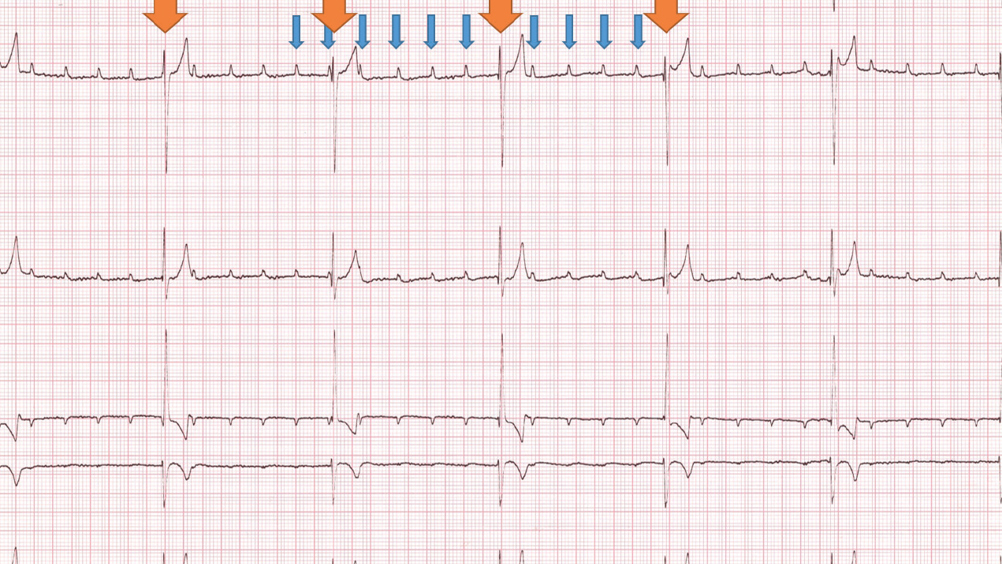
This case report describes the patient journey of a young Cockapoo with symptomatic bradycardia, from admission to a referral hospital, investigations and management with pacemaker implantation, until discharge from the hospital. The case describes the general physical examination findings specific to symptomatic bradycardia, as well as common investigative tests performed in cardiology cases such as indirect blood pressure measurement, biomarkers (in-house cardiac troponin I), six-lead electrocardiography and comprehensive echocardiography. This case also describes the specialist nursing role during pacemaker implantation, including pacemaker programming using telemetry, the use of fluoroscopy with a C-arm, and surgical pull list and theatre set up. The post-surgical follow-up and further optimisation of the pacemaker settings is also described. Third degree atrioventricular block is the most common reason for pacemaker implantation. Awareness of the patient journey during pacemaker implantation is important to provide adequate support and advice to owners of canine patients with symptomatic bradycardia before referral.
Lucy was a 3 year and 2 month old, female neutered Cockapoo. The chief presenting complaint was lethargy and exercise intolerance associated with severe bradycardia at 30 beats per minute (bpm) noted by the referring veterinary surgeon.
Lucy presented to the cardiology department for a consultation. Lucy had been in the owner’s possession since she was 6 months old and was the only pet in the house. She was fully vaccinated and up to date on worming and flea/tick treatment. No coughing or sneezing had been noted. Her urination and defecation were normal, and no vomiting was observed.
Approximately 2 days before presentation, Lucy became lethargic and reluctant to exercise. Her appetite was reduced. Her referring veterinarian noted a bradycardia at 30 bpm. No syncopal or collapsing episodes were reported by the owners.
Lucy presented as quiet, alert and responsive with a weight of 13 kg and a body condition score of 6/9. Her mucus membranes were pink and moist with a capillary refill time of less than 2 seconds. Lymph nodes were all within normal limits. The abdomen was not tense or painful and no abnormalities were detected via palpation. Respiratory rate was 32 breaths per minute with normal effort and clear lung sounds were heard on auscultation. Her heart rate was 40 bpm with no murmur present. Lucy’s pulses were strong and no deficits were felt. Her rectal temperature was 37.8°C.
Register now to continue reading
Thank you for visiting The Veterinary Nurse and reading some of our peer-reviewed content for veterinary professionals. To continue reading this article, please register today.

