References

Aspinall V. The Complete Textbook of Veterinary Nursing, 2nd Edition. London: Saunders Elsevier; 2011

Chan DL. Refeeding syndrome in small animals. In: Chan DL Chichester: John Wiley & Sons Ltd; 2015

BSAVA Textbook of Veterinary Nursing, 5th Edition. In: Cooper B, Mullineaux E, Turner L Gloucester: BSAVA; 2011

Firth A. Early enteral nutrition—principles and practice. The Veterinary Nurse. 2013; 4:(7)392-9 https://doi.org/10.12968/vetn.2013.4.7.392

Freeman L, Becvarova I, Cave N WSAVA nutritional assessment guidelines. J Feline Med Surg.. 2011; 13:(7)516-25 https://doi.org/10.1016/j.jfms.2011.05.009

Gray K. Management of tubes, lines and drains. The Veterinary Nurse. 2018/2019; 9:(10)525-31 https://doi.org/10.12968/vetn.2018.9.10.525

Hodshon B, Tobias KM. Esophagostomy feeding tubes. Clinician's Brief. 2014; 12:(2)66-72

Lumbis RH. How to place commonly used feeding tubes in dogs and cats. The Veterinary Nurse. 2017; 8:(2)104-15 https://doi.org/10.12968/vetn.2017.8.2.104

Orpet H, Welsh P The Handbook of Veterinary Nursing, 2nd Edition. Oxford: Wiley-Blackwell; 2011

Parker VJ. Nutritional management of hospitalised dogs and cats. The Veterinary Nurse. 2013; 4:(8)478-85 https://doi.org/10.12968/vetn.2013.4.8.478

Parker VJ, Freeman LM. Comparison of various solutions to dissolve critical care diet clots. J Vet Emerg Crit Care (San Antonio). 2013; 23:(3)344-347 https://doi.org/10.1111/vec.12047

Ross S. Utilization of Feeding Tubes in the Management of Feline Chronic Kidney Disease. Vet Clin North Am Small Anim Pract.. 2016; 46:(6)1099-114 https://doi.org/10.1016/j.cvsm.2016.06.014

Smith AJ. Perspectives on Feeding and Nutrition. Vet Clin North Am Small Anim Pract.. 2019; 49:(3)501-17 https://doi.org/10.1016/j.cvsm.2019.01.014

van Schoor M. How to manage indwelling feeding tubes in critically ill dogs and cats. The Veterinary Nurse. 2015; 6:(2)118-23 https://doi.org/10.12968/vetn.2015.6.2.118

How to tube feed

02 May 2019
13 mins read
Volume 10 · Issue 4

Abstract

There are many different enteral feeding tubes available for use in veterinary practice to support the nutritional management of a wide variety of patients. This article aims to provide practical advice and guidance with regards to all stages of tube feed administration to ensure it is done safely and the tube is of maximum benefit to the patient and its recovery from illness or injury.

Freeman et al (2011), in the WSAVA nutrition guidelines, described nutrition as the fifth vital sign because adequate nutrition is vital to the healing, health and wellbeing of all animals. There are many indications for enteral feeding of veterinary patients due to many injuries and diseases resulting in a patient being unwilling or unable to ingest adequate calories to maintain health. Examples include mechanical or physical problems such as maxillofacial trauma following road traffic collisions, primary gastrointestinal problems affecting nutrient absorption, or anorexia as a result of metabolic derangements (Ross, 2016). Smith (2019) also highlighted the fact that many patients fail to eat enough food towards the end of their life for various reasons, including patients with conditions such as chronic kidney disease. Whatever the underlying reason for the malnourishment, feeding tubes are an excellent intervention to help support such patients, and it is vital that all veterinary professionals are comfortable with their use to ensure maximum patient benefits.

Nutritional requirements

All patients receiving enteral nutrition should have a nutritional requirement calculation completed and reviewed daily, and it is ideal to record this on a nutrition calculation chart to refer back to. It is recommended to calculate their resting energy requirement (RER) in kilocalories per day (kcal/day) and aim to fulfil this while hospitalised as they are expending minimal energy while restricted to a cage. Veterinary professionals should check the RER has been calculated before going ahead with the next feed.

An accurate bodyweight (BW) in kilograms (kg) is required to calculate the RER, and the linear formula of (BW x 30) + 70 = kcal/day is a reasonable approximation for patient requirements when they weigh between 3 and 25 kg (Firth, 2013; Parker, 2013; van Schoor, 2015). It is preferable to use the allometric, or exponential formula to calculate RER for all patients as it considers the patient's body surface area and metabolic rate: 70 x (BW)0.75 = kcal/day (Firth, 2013; Parker, 2013; van Schoor, 2015). This latter formula is recommended for patients that are less than 3 kg and greater than 45 kg because the former tends to overestimate the patient's requirements (Firth, 2013).

Parker (2013) advised to use the patient's current weight for these RER calculations, however if they are very obese it is wise to alter this and use a BW closer to their ideal weight to avoid overfeeding. When the patient with a feeding tube in situ is discharged home and is more active, the RER can be multiplied by an appropriate factor to identify their maintenance energy requirements (MER); this factor typically ranges from 0.8 to 1.8 and the factor used should be discussed with the veterinary surgeon (VS).

Why an enteral nutrition and feeding plan?

Before enterally feeding, if a veterinary professional is not familiar with the patient they should review the patient's records to learn a little more about them and establish why the patient requires enteral feeding. There should be evidence in their records of nutritional screenings having been completed, which includes consideration of their life stage, BW, body condition score (BCS) and muscle condition score (MCS) (Freeman et al, 2011; Parker, 2013). Knowledge of patients and their background is essential to the provision of quality care and management.

Their nutritional plan should be checked to establish how many days the patient has been receiving enteral nutrition for thus far, and therefore how much food per feed needs to be given at that moment in time. Hodshon and Tobias (2014) recommended splitting the daily RER into 4 to 6 feeds according to estimated stomach capacities. When a patient has been anorexic or hyporexic for a period of time their stomach capacity will reduce, so this has to be taken into consideration, and refeeding syndrome needs to be avoided. Hodshon and Tobias (2014) indicated a gastric capacity of 5 to 10 ml/kg should be assumed during food reintroduction, reaching a normal 45 to 90 ml/kg capacity when realimentation is complete.

Chan (2015) recommended providing up to 20% of their RER on day 1, rising incrementally over the next 4 to 10 days in accordance with their progress, which should be closely monitored. van Schoor (2015) recommended providing 25 to 50% of the calculated RER on day 1, gradually increasing the amount over a few days according to their progress. Firth (2013) recommended for debilitated patients, who cannot tolerate immediate feeding with liquid or blenderised foodstuffs, administering 0.5 to 0.5 ml/kg of an isotonic solution containing sodium, glucose and key amino acids, either intermittently or by constant rate infusion (CRI), increasing the volume by 50% every 8 to 12 hours depending on the progress the patient makes and their tolerance of the feeds. Firth (2013) then advised that the volume and caloric content of the food can be increased on day 2 or 3 according to patient progress, until 100% of the RER is eventually being fed daily. Ultimately, the period over which food is reintroduced is at the discretion of the VS, who should discuss their plans with all caregivers.

Pre-feed patient checks

Prior to administering the next feed, the patient's records should be checked to ascertain how well they have tolerated recent feeds. The veterinary professional should be looking for information regarding any problems that have been encountered associated with the actual feeding tube itself, or the patient's response to the feed. If any of this information is ambiguous on the records, clarity should be sought from those members of staff who have previously been responsible for the patient.

Veterinary professionals should double check the diet to be fed, the route of administration, the amount required, and the frequency (Parker, 2013). They should also assess for themselves whether the patient is haemodynamically stable enough to receive the next feed, or discuss this with a senior member of staff if they are unsure. The patient's cardiovascular stability should be assessed, their body temperature checked, recent electrolyte levels reviewed, their acid–base balance considered, and any recent episodes of vomiting or regurgitation should be noted. Parker (2013) and Lumbis (2017) both discussed that assessing these parameters is good practice to establish whether there are any clinical signs of refeeding syndrome, and if there are any abnormalities these should be corrected prior to feeding. It may have been some time since the last blood testing occurred, so bringing this to the attention of the case VS would be appropriate prior to feeding.

Once these patient checks are complete and it has been established the patient is able to receive the next feed, the veterinary professional undertaking the feed should assess whether they are going to need any assistance with restraint during the process, and feeding a patient in sternal recumbency is encouraged as this is a more natural feeding position (Lumbis, 2017).

Foodstuff preparation

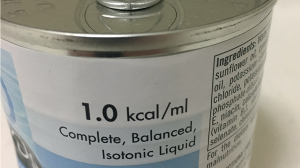
The first thing to do when preparing to feed a patient is of course to select the correct food-stuff, as per the patient's records, which may be a pre-prepared product or may involve mixing a solution together. Some practices like to feed the patients a more liquid solution and others like to blenderise normal foodstuffs; Lumbis (2017) advised that if foodstuffs are being blenderised it is wise to strain them prior to administration. Whatever product is being used, the kcal per millilitre (ml) of food will typically have been used in the nutritional calculations to determine how many ml are needed per feed to meet the patient's requirements, however there are alternative ways to determine food requirements. The kcal/ml of the foodstuff selected should be double checked against the patient's records just to ensure it is definitely the right product prescribed by the VS (Figures 1a and 1b).

Figure 1a. This foodstuff is calorie-dense, providing 1 kcal/ml.
Figure 1b. This foodstuff is also calorie-dense but presents the information in a different manner, as metabolisable energy. There are 380 kcal in 375 ml which equates to fractionally more than 1 kcal/ml.

The correct volume of food required should be drawn up into appropriately sized syringes (Figure 2), and it is recommended to warm these up to body temperature. This can be achieved by putting the syringes in a bowl of warm water for a period of time in advance of the scheduled feed. Sterile water should be drawn up for the pre-feed flush in case the tube has migrated into the trachea and the syringe kept sterile prior to use, either by replacing this back in the syringe packet or by attaching a sterile needle to the syringe (Figures 3a and 3b). Sterile or tap water can be used for the post-feed flush, and it is ideal to warm all of the water up to body temperature too. It is often easier to draw the total flush volume up into one syringe from the sterile supply as this is more time-efficient.

Figure 2. This shows some liquid diet which has been drawn up and warmed ready for administration.
Figure 3a. This shows the sterile pre-flush saline drawn up and being kept sterile by reinserting it into its original packaging.
Figure 3b. This shows the sterile pre-flush saline drawn up and being kept sterile by attaching a sterile needle.

The patient's record should also be checked at this point to establish whether any medications need to be administered down the feeding tube at the same time as the food, and these should be prepared accordingly (Figure 4).

Figure 4. This shows two syringes of medications (top) which have also been drawn up ready to be administered alongside the feed.

Pre-feed tube checks

While the food and flush is warming up the veterinary professional can don some gloves and start checking the feeding tube itself. Its integrity needs to be assessed, the security needs to be checked, and all connectors should be checked for damage. The person doing these checks should also ensure they are familiar with that particular tube, how it should be used, and what type of foodstuff is suitable to administer through it — is it a nasogastric (NG) tube, nasooesophageal (NO) tube, oesophagostomy (O) tube, gastrostomy (G) tube or a type of enterostomy tube? There are a wide range of different styles of feeding tubes available from different manufacturers (Figures 5a, b, c, d), so it is wise to check how they work prior to commencing the feed, and ask for help if there is any uncertainty. Lumbis (2017) advised that feeding tubes should also be clearly labelled as such to avoid them being confused with an intravenous line, for example.

Figure 5a. This is an example of a simple feeding tube.
Figure 5b. This is also an example of a simple feeding tube but note the slightly more complex end to the tube when compared with that of Figure 5a.
Figure 5c. This is an example of a wider bore feeding tube. This one comes with luer lock syringes that are designed to be used with it.
Figure 5d. This is an example of a feeding tube where an adaptor would be required to facilitate the attachment of normal sized luer syringe hubs; otherwise dosing syringe tips will fit in the end.

The insertion or stoma site (Figure 6) and associated dressings should be checked at this point too, and the records checked regarding whether a clean and dressing change is required; dressing should be replaced at least once a day (Lumbis, 2017). If a feeding tube has just been placed then a radiograph is typically taken to confirm its placement, or equipment such as a capnograph could be used to check an NG or NO tube is not in the trachea (Gray, 2019). Gray (2019) also suggested aspirating from an NG tube to identify gastric juices is a useful method to check placement (Figure 7). Only once all of the above checks have been made should a feed be administered.

Figure 6. This shows an oesophagostomy (O) tube in situ. Stomas should be checked along with the security of the finger trap suture.
Figure 7. This puppy has an nasogastric (NG) tube, and these can be gently aspirated prior to flushing and food administration to confirm their position and or to remove some of the gastric juices that may have built up since the last feed.

Flushing and feeding

Lumbis (2017) advised that encouraging the patient to feed naturally should be done before all tube feeds to check whether they have regained their appetite or not, however any contraindications to this must be discussed with the VS first. Prior to the food being administered the feeding tube should be flushed with sterile water; this is to check for both patency and position. If it has migrated into the trachea the sterile water will elicit a cough response, but not introduce pathogens into the tract.

There are varying volumes recommended in literature for this purpose, and seemingly no definitive answers relating to the volume. Orpet and Welsh (2011) advised 5 ml, Aspinall (2011) 2 ml, and Cooper et al (2011) 5 ml; van Schoor (2015) recommended 5 to 20 ml depending on the size of the animal and the size and length of the tube itself, Hodshon and Tobias (2014) advised 10 to 20 ml, whereas Lumbis (2017) recommended 5 ml. Some manufacturers include instructions for use with the feeding tubes they supply, recommending volumes for flushing, so these should be taken into consideration too.

Ultimately, enough water needs to be administered to completely flush through the entire length of the tube to ensure it is clear of debris, and enough water needs to be administered to elicit a cough response should the tube have migrated. It would be advisable for all veterinary professionals to check the tubes they commonly use and test how much fluid is required to achieve these objectives. Van Schoor (2015) also warned about considering flush volumes being administered when calculating a patient's total daily fluid requirement to avoid fluid overload.

The author recommends slowly administering a small volume of the sterile pre-feed flush initially, for example 0.5 to 1 ml, to ensure only a small quantity has been delivered should the tube have migrated into the patient's trachea. If there is no cough response, and therefore confidence that it is in the gastrointestinal tract, then the rest of the desired pre-feed flush volume can be administered a little more rapidly. For example, if 5 ml pre-flush has been drawn up and 1 ml has been given very slowly and there is no cough response, the other 4 ml could be safely given over 1 minute.

Following the pre-feed flush, the food can then be administered. Again there does not seem to be clear consensus in literature regarding the speed at which this should be administered. The author recommends, from experience, that a feed takes between 10 and 20 minutes to administer (or even longer for very large volumes), so for a volume of 10 ml this would equate to 1ml per minute over 10 minutes; this is slow enough to avoid problems such as regurgitation and has proven effective experientially. Administering a small increment, allowing the patient to receive this, and observing for any adverse reactions is recommended before then giving the next increment. Orpet and Welsh (2011) advised that larger syringes, for example 20 ml syringes or larger, require less force to be used to expel the contents during a feed, so can help with control of delivery and prevent rupture of the tube (Figure 8).

Figure 8. This shows a patient being fed via their oesophagostomy (O) tube.

Following the feed the tube needs to be flushed again to clear it of any remaining food-stuffs that may harden and cause a blockage (Figure 9). There is no requirement for this to be sterile water as the tube position has already been confirmed by the pre-feed flush. As per the pre-feed flush discussion, the recommended volume for this second flush also varies in the literature and there is no consensus around one specific volume. Cooper et al (2011) and Lumbis (2017) recommended a 10 ml flush following liquid diets and 20ml following blenderised diets, due to blockages being more likely with the latter. Van Schoor (2015) advised 5 to 20 ml should be used, again depending on the size of the patient and tube capacity, and different manufacturers will recommend specific volumes for their own tubes if they provide product information of this nature. As for the pre-feed flush volume, it is advised that all veterinary professionals check the volume required to clear the tubes they are using in their practices so at least the correct minimum volume is being used to clear the length of the tube.

Figure 9. This picture shows an oesophagostomy (O) tube in a cat. This feeding tube had not been used for approximately a couple of days as the cat has started eating voluntarily, so was about to be removed, however a lump of dried food can be seen within the lumen as an example of a blockage we should try to avoid (dark spot about 1 inch to the right of the end of the suture).

Ultimately, enough water needs to be administered to ensure all food debris is cleared from the tube's lumen and the author typically uses around 10 ml for this purpose, which has always been deemed sufficient experientially. This water, as previously mentioned, should also be warmed to body temperature and not given so fast that it results in regurgitation.

Recording

Once a feed is complete it needs to be recorded on the patient's hospitalisation chart and/or clinical records. The volume of pre-feed flush and post-feed flush should be recorded, alongside the volume of food administered. If it is not already documented, the type of foodstuff used should also be recorded. There needs to be space on the hospitalisation chart to record how well the tube feed was tolerated by the patient, whether there were any noteworthy problems during the process, and what interventions were performed to rectify any issues. It should also be recorded when dressings have been removed and replaced, and any relevant comments about the insertion site or stoma (Figure 10). There is a toolkit available associated with the WSAVA guidelines which contains a chart for documenting nutritional intake, and this can be downloaded for free from the WSAVA website.

Figure 10. This shows a new, sterile primary dressing around the stoma site. Note that gloves should be worn at all stages of the process of tube feeding, and any time the tube is being checked and dressings changed.

If complications are encountered during the process of feeding the intervention should cease, the cap be replaced on the tube and the tube secured appropriately, and the VS in charge of the case informed. The problem can be investigated further and changes to the nutritional plan made accordingly, which may involve the complete replacement of the tube for example, or performing a clinical examination of the patient, or interventions being implemented to unblock a tube if that is the suspected problem. It is worth noting at this point that Parker and Freeman's (2013) study, which investigated the efficacy of different solutions to dissolve critical care food clots, found that water was significantly better (p<0.001) than all carbonated beverages and cranberry juice at dissolving the clots, however the best solution was a mixture of ¼ teaspoon pancreatic enzymes and 325 mg sodium bicarbonate in 5 ml of water. This study was not performed using actual feeding tubes in live patients that had blocked, so the authors recommended future in vivo studies to corroborate their findings, however it does provide confidence that water, which is readily available, has potential to be effective at unblocking feeding tubes and carbonated beverages which may cause digestive upset can be avoided.

The patient's BW should be recorded at least daily, ideally twice a day, to ensure their caloric intake is adequate, and monitoring of their overall clinical condition is essential to judge the efficacy of the nutritional plan as a whole.

Conclusions

The process of administering food via a feeding tube may seem like a fairly straightforward task in veterinary practices where they are routinely used, however there are many factors to take into consideration to detect and prevent problems from occurring. It is the responsibility of all veterinary professionals to ensure they are familiar with the feeding tubes that have been placed in the patients they are nursing, and that they have read all of the manufacturer's instructions if these have been supplied. It is advisable for all practices to have a protocol in place relating to the use and management of feeding tubes to ensure their longevity in terms of functionality, and protocols are very useful as a training tool for less experienced members of the team.

KEY POINTS

  • Feeding tubes are an excellent way to ensure a patient receives adequate nutrition in situations where they may be unwilling or unable to eat for themselves, and nutritional requirements should always be carefully calculated.
  • There are a variety of feeding tubes available for use in veterinary patients, and all clinical staff members should be appropriately trained in their use.
  • It is essential to administer a sterile pre-feed flush to ensure the patency and position of the feeding tube.
  • Foodstuffs should always be warmed to body temperature and fed slowly to avoid problems such as regurgitation.
  • Feeding tubes should be flushed with an absolute minimum of 5 ml of water following every feed to ensure the lumen is patent and free of debris.