References

Alveraz A. Treatment of cranial cruciate ligament rupture in dogs - an overview. Veterinary Focus. 2011; 21:(2)39-46

Brandsson S, Faxén E, Kartus J, Eriksson BI, Karlsson J. Is a knee brace advantageous after anterior cruciate ligament surgery?. Scand J Med Sci Sports. 2001; 11:(2)110-4

Carr BJ, Canapp SO, Meilleur S, Christopher SA, Collins J, Cox C. The Use of Canine Stifle Orthotics for Cranial Cruciate Ligament Insufficiency. Veterinary Evidence. 2016; 1:(1) https://doi.org/10.18849/ve.v1i1.10

Comerford E, Forster K, Gorton K, Maddox T. Management of cranial cruciate ligament rupture in small dogs: a questionnaire study. Vet Comp Orthop Traumatol.. 2013; 26:(6)493-7 https://doi.org/10.3415/VCOT-12-06-0070

Corr S. Decision making in the management cruciate disease in dogs. In Practice. 2009; 31:164-71

Davidson JR, Kerwin SC, Millis DL. Rehabilitation for the Orthopedic Patient. Vet Clin North Am Small Anim Pract.. 2005; 35:(6)1357-88

Davis A. Tibial Plateau Levelling Osteotomy (TPLO). Veterinary Nursing Journal. 2009; 24:(3)27-9

Di Dona F, Della Valle G, Fatone G. Patellar luxation in dogs. Vet Med (Auckl).. 2018; 9:23-32 https://doi.org/10.2147/VMRR.S142545

Harasen G. Making sense of cranial cruciate ligament disease Part 1: Epidemiology and Pathophysiology. UK Vet Companion Animal. 2011; 16:(1)29-32

Morris E, Lipowitz AJ Comparison of tibial plateau angles in dogs with and without cranial cruciate ligament injuries. J Am Vet Med Assoc.. 2001; 218:(3)363-6

Millis DL, Levine D. Canine Rehabilitation and Physical Therapy.: Elsevier; 2014

Norris S. Referral nursing – cranial cruciate ligament rupture in dogs. Veterinary Nursing Journal. 2012; 27:(3)91-115

Shaw KK. Physical rehabilitation for canine patients post cranial cruciate ligament surgery. Companion Animal.. 2017; 22:(12)714-22

Vasseur PD. Clinical results following nonoperative management for rupture of the cranial cruciate ligament in dogs. Vet Surg.. 1984; 13:243-6

von Pfeil D, Kowaleski M, Glasman M, Dejardin LM. Rogers - von Pfeil. Nonsurgical treatment for cranial cruciate ligament rupture; Letter to the Editor. JAVMA18. JAVMA.. 2018; 253:1241-42

Whitehair JG, Vasseur PB, Willits NH. Epidemiology of cranial cruciate ligament rupture in dogs. J Am Vet Med Assoc.. 1993; 203:(7)1016-9

Cruciate ligament disease and the role of the Balto Knee Brace
02 September 2019
17 mins read
Volume 10 · Issue 7

Abstract

Cruciate disease is the most prevalent cause of hindlimb lameness amongst the canine population. As a result of the prevalent nature of the condition, improved management strategies are continually being sought from both a surgical and conservative perspective. This article discusses cruciate disease, and the KVP ‘Balto’ brace in relation to its use as part of conservative management strategy.

Cruciate ligament disease, a term developed to include both acute ruptures and the more common chronic degeneration of the cranial cruciate ligament (CCL) (Corr, 2009), is the most common cause of hindlimb lameness in the dog (Harasen, 2011). With a likelihood of rupture of the CCL in the contralateral limb estimated to be approaching 60% (Harasen, 2011), and the inevitable development of degenerative joint disease in the affected stifle, cruciate disease can have a huge impact on animal wellness and quality of life. New techniques for repair and/or management are continually being sought in an attempt to address this issue. This article discusses the background of the condition and different management techniques available for treatment, including use of a the Balto Knee Brace for support, designed and manufactured by JOYVET Italy and supplied by KVP International.

Background

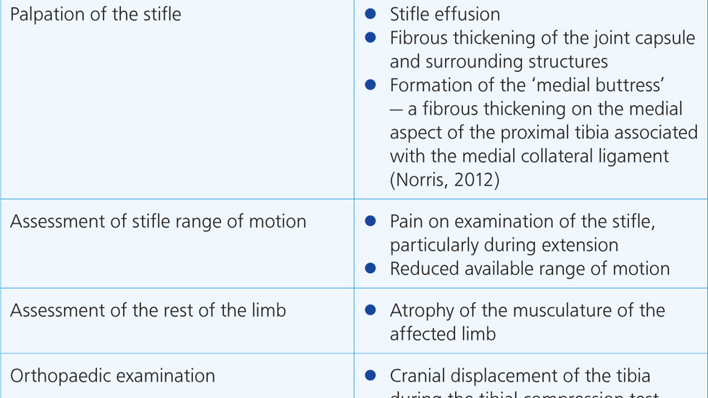
The role of the cranial (alongside the caudal) cruciate ligament, which originates from within the intercondylar notch of the distal femur and inserts on the intercondylar area of the tibial plateau, is to provide support to the stifle and prevent tibial thrust — movement of the tibia in a cranial direction (Alveraz, 2011). Clinical signs therefore arise from loss of this function and include one or more of the findings listed in Table 1 on assessment of the patient.


Table 1. Findings during an examination of patients affected by cruciate disease
Type of assessment Clinical findings
Gait and postural assessment
  • A varying degree of lameness
  • Off-loading the affected limb during stance
  • An altered sit position whereby the stifle and lower limb are extended laterally (Shaw, 2017)
Palpation of the stifle
  • Stifle effusion
  • Fibrous thickening of the joint capsule and surrounding structures
  • Formation of the ‘medial buttress’ — a fibrous thickening on the medial aspect of the proximal tibia associated with the medial collateral ligament (Norris, 2012)
Assessment of stifle range of motion
  • Pain on examination of the stifle, particularly during extension
  • Reduced available range of motion
Assessment of the rest of the limb
  • Atrophy of the musculature of the affected limb
Orthopaedic examination
  • Cranial displacement of the tibia during the tibial compression test
  • Cranial displacement of the tibia during the cranial drawer test
Diagnostic imaging
  • Stifle effusion
  • Loss or cranial displacement of the intrapatellar fat pad
  • Osteophyte formation on the femoral condylar ridges
  • Development of secondary degenerative joint disease (Harasen, 2011)

Genetics and predisposing factors

There are several risk factors which increase the likelihood of the development of cruciate disease. In an extensive study on predisposing factors, Rottweilers, Newfoundlands, and Staffordshire Terriers had the highest prevalence of CCL rupture, whereas Dachshunds, Basset Hounds, and Old English Sheepdogs had the lowest; demonstrating that genetics have an influence on development of the disease. The same study demonstrated that neutered males and females had a higher prevalence of rupture than intact dogs, and that dogs weighing more than 22 kg had a higher prevalence than those weighing less than 22 kg (Whitehair et al, 1993). More recent research also adds Labrador Retrievers and Golden Retrievers to the list of breeds which are over-represented, among others (Harasen, 2011).

Environmental and lifestyle factors including obesity and the feeding of high-energy diets have also been shown to increase the incidence of cruciate disease (Harasen, 2011).

Due to stifle instability and internal rotation of the tibia, dogs with a grade 3 or 4 patellar luxation have been shown to have an increased likelihood of developing cruciate disease (Harasen, 2011), with one study demonstrating that 41% of dogs with medial patellar luxation experienced concurrent cruciate rupture. Given the frequent nature of patellar luxation amongst small dog populations in particular, the condition is an important factor in the incidence of the cruciate disease (Di Dona et al, 2018).

Much interest has been given to the angle of the tibial plateau, and both its impact on the likelihood of the development of cruciate disease, and also its adjustment as a surgical technique used to treat dogs with cruciate rupture procedures such as the tibial-plateau-leveling osteotomy (TPLO) and tibial tuberosity advancement (TTA). It has been demonstrated that, when measured, dogs diagnosed with cruciate disease had a higher tibial plateau angle than those without (Morris and Lipowitz, 2001). With this in mind, it would be an extremely useful development within the veterinary profession if both animals which are high risk due to predisposing factors (such as breed, bodyweight, history of cruciate rupture in the con-tralateral limb), and those that have suffered a rupture could be screened to enable risk reducing measures to be undertaken (such as weight reduction), and to allow for selection of the best treatment option for that individual patient.

Management of cruciate disease

Management of cruciate disease currently falls into two broad categories: surgical or non-surgical.

Surgical management

Surgical management is recommended as the preferred treatment option for many diagnosed patients, particularly those weighing more than 15 kg or with concurrent meniscal injury. The goals of surgical management are to achieve stabilisation of the stifle, while also reducing pain and facilitating return to function for the patient. A desirable outcome is also to minimise the progression of secondary degenerative joint disease, and to assess the meniscus and remove any damaged tissue (Shaw, 2017).

The currently available surgical techniques fall into three broad categories: intracapsular, extracapuslar and orthotomy procedures. Three of the most commonly used procedures in practice today include the lateral suture technique (extracapsular), and the TTA and TPLO (orthotomy techniques) (von Pfeil et al, 2018). All three techniques allow visualisation and, if necessary, removal of damaged meniscal tissue (Davis, 2009). The goal of both the TTA and TPLO techniques is to eliminate ‘cranial tibial thrust’ — the cranial movement of the tibias relative to the femur. This is achieved by incising and rotating the proximal tibia, then securing it in place with a bone plate in the case of the TPLO; and by transposing the tibial tuberosity cranially to shift the alignment of the patellar tendon, which is secured in place by a titanium cage and bone plate in the case of the TTA (Millis and Levine, 2014).

Non-surgical management

Non-surgical (or conservative) management generally consists of some or all of the following: use of non-steroidal anti-inflammatory drugs, weight management, exercise modification often including cage rest initially, stifle support in the form of a brace, and physical therapy such as physiotherapy and/or hydrotherapy (Comerford et al, 2013). The goals of non-surgical management are again to address pain and allow a return to function for the patient.

It has been shown that this form of management is often best suited to dogs weighing less than 15 kg, where a success rate of 85% was demonstrated (Vasseur, 1984); and those without concurrent meniscal injury — which is estimated to affect around 45% of patients (Davidson et al, 2005). It remains the only treatment option for patients for which general anaesthesia or surgery is not a desirable option, such as those diagnosed with co-morbidities.

Stifle support

Much interest has been given recently to the use of stifle support in the form of a brace for both cruciate rupture patients that are undergoing conservative management; and in the immediate postoperative period for those that have undergone surgical management.

Human studies have demonstrated positive results in terms of a reduction in postoperative pain scores, and a decrease in postoperative complications in patients which have undergone anterior cruciate surgery and wore a supportive brace for 3 weeks following surgery, compared with those who underwent the same procedure and did not wear a brace (Brandsson et al, 2002).

Given these encouraging results in human patients, research has been carried out to determine whether the canine patient would benefit from bracing the stifle during conservative management of cruciate disease. A small study of ten dogs with unilateral cruciate rupture conducted in 2016 indicated that use of a custom stifle orthotic improved weight bearing through the affected limb (Carr et al, 2016). The study was limited by the small sample size and lack of control group, but the results are encouraging and so further studies are warranted.

The Balto Knee Brace has been developed by JOYVET for such purposes. Features of the brace include removable alu-minium splints to restrict movement within the stifle, and an anchor strap and abdominal strap to ensure the brace is well secured. It is manufactured using breathable fabric to minimise irritation, and the straps are adjustable and can be trimmed to fit each individual patient to maximise comfort levels. Other proposed uses include use in contralateral limb support and in patients with other conditions affecting the stifle including luxating patella, arthritis, or arthrosis (KVP International, 2019). A number of case studies are presented here written both by the author, and contributed from veterinary practices.

Case report 1 — Bradley

History

An 8-year-old male entire Boxer presented for a physiotherapy assessment following a diagnosis of cruciate rupture to the left hindlimb around 4 weeks prior to this appointment by the referring veterinary surgeon. General anaesthesia for radiographs/surgery was opted against as the dog was an unstable epileptic, and so conservative management of the condition was being sought.

Clinical examination and diagnosis

Examination during the initial assessment revealed the following findings and were indeed indicative of cruciate rupture:

  • 9/10 lame left hindlimb (toe-touching only)
  • Marked atrophy of the left hind quarter (girth measuring 33 cm at widest point vs 41 cm right)
  • Effusion of the left stifle
  • Palpable medial buttress
  • Positive cranial drawer.

Treatment plan

Discussion was had with the dog's owner regarding management of cruciate disease. As the dog was over 15 kg it was advised that surgical management was likely to have the most favourable outcome; but due to the dog's unstable epilepsy the owner felt this was not a viable option. A treatment plan for conservative management was discussed with the owner to include the following:

  • Continued use of meloxicam as prescribed by the referring veterinary surgeon
  • Maintenace of bodyweight (the dog was in ideal body condition at the time of examination)
  • Limiting exercise to a maximum of 15 minutes per walk on the leash as the dog was currently being exercised together with the other dogs off-leash for up to 1 hour once or twice daily
  • Rehabilitation sessions consisting of land-based physiotherapy including weaves, cavaletti poles, exercises on the ramp and gentle use of the balance board; hydrotherapy in the underwater treadmill; and electrotherapy were advised initially twice weekly. This was to address muscle atrophy and promote limb use, enhance proprioception and assist with pain management, and would also include a home programme of exercises for the owners to complete
  • Use of the Balto Knee Brace without aluminium splints for stifle support during walks and physiotherapy sessions in clinic and at home, which was fitted in the clinic, and demonstrated to the owners.
Figure 1. Bradley, an 8-year-old Boxer, presented following a diagnosis of cruciate rupture. Due to unstable epilepsy his owners opted for conservative management using the Balto Knee Brace.

Response to treatment

The dog responded well to the conservative management plan. The owner was able to bring the dog for twice weekly rehabilitation sessions for 3 weeks, after which point they could bring him once weekly.

After two sessions the owner reported that the brace was rubbing the undercarriage of the dog causing some redness of the skin which may have occurred as result of the dog being relatively hairless in that region. An alternative brace (the Ligatek prototype with range of motion) was ordered to ensure the dog did not develop secondary skin issues related to use of the brace.

After six rehabilitation sessions on a twice-weekly basis it was noted that limb use, foot placement and muscle tone were already beginning to improve. The dog was described as weight bearing more evenly through the hindlimbs.

Just over a month after initiating conservative management, the muscle mass of the left hindlimb was seen to be improving (by 2 cm in measured girth), weight bearing through the left hindlimb was also improving (the lameness was now graded 3/10) and the dog appeared comfortable on flexion and extension of the stifle. Given the marked atrophy of the limb it was likely that the dog would require long-term management to address this, but both the owner and the rehabilitation therapist were impressed by the dog's progress so far.

Discussion

This case describes how a conservative management plan can be used as an alternative to surgery where the latter is contraindicated in dogs suffering from a ruptured CCL.

The dog's owners both agreed that although they had initially been concerned that the brace could be difficult to fit at home, it did not take long for them to get used to it, and they acknowledged that the video footage they were supplied with the brace was helpful in making this process easier. The brace was well tolerated by the dog, and used by the owners while walking him, completing the home exercise programme and during participation in the land-based physiotherapy in the clinic.

The patient still had a long journey ahead in terms of progress but his improvement over the first month following initiation of rehabilitation was very encouraging.

Case report 2 — Max

History

A 10-year-old Springer Spaniel presented for an ongoing acupuncture session to assist with pain management of osteoarthritis of the left stifle which was thought to have occurred secondary to a cruciate rupture 2 years previously, which was treated conservatively (Figure 2). The dog's management plan currently consisted of the following:

  • Analgesics prescribed by the referring veterinary surgeon including meloxicam, tramadol and gabapentin
  • Exercise modification whereby the owner would avoid allowing the dog to engage in rough-play with other dogs and would avoid ball-throwing as both of these activities had resulted in a flare-up of the dog's symptoms in the past. This was frustrating for the dog and owner as the dog was still energetic and ‘looked for’ more exercise, according to the owner
  • Acupuncture sessions as required when the owner perceived the dog was more painful.
Figure 2. Max, an 10-year-old Springer Spaniel, presented with pain as a result of osteoarthritis, and a resultant reduced ability to exercise. A Balto Knee Brace was supplied for use during exercise.

Examination

On clinical examination prior to administering acupuncture the following findings were noted:

  • The dog was 3/10 lame on the left hindlimb
  • The left stifle was thickened with a reduced range of motion
  • There was moderate atrophy of the musculature of the left hindquarter.

The owner was happy with the dog's pain control at present but expressed frustration (not for the first time) that he felt unable to increase the dog's exercise as he was concerned this would result in an exacerbation of the dog's symptoms. The dog was currently being exercised for 20 minutes on the leash twice daily.

Treatment

The dog tolerated the acupuncture extremely well, and was comfortable and relaxed during the session. Discussion was had with the owner regarding the dog's exercise in attempt to reduce frustration. The owner was offered the option of trying the Balto Knee Brace for the dog to wear during exercise, to monitor whether applying this additional support to the affected stifle would enable to dog to participate in more exercise without any impact on the dog's condition. It was agreed that the owner would continue to avoid high impact exercise for the dog, such as ball chasing and rough play, but could gradually increase the duration of the on-leash exercise by 5 minutes per walk per week up to a maximum of 35 minutes twice daily until the dog was reassessed. Food puzzles were also suggested as an additional way to occupy the dog and address any frustration.

Response to treatment

The dog presented for acupuncture again 3 weeks later. His clinical signs on examination were unchanged from the previous visit. History taking from the owner revealed that the owner had found the brace difficult to put on the dog initially, and that he had to rely on times where his wife was available to assist with holding the dog. The dog had tolerated wearing the brace extremely well, and there were no issues relating to it staying in place during exercise.

The owner had successfully managed to increase the exercise to 35 minutes twice daily without the development of any obvious lameness, or any behavioural change relating to pain. He felt as a result of this the dog was calmer in the house between walks. He had also introduced food puzzles for the dog to play with between walks.

Discussion

This case study describes how a brace when used for stifle support may improve comfort levels and tolerance relating to exercise in patients suffering from osteoarthritis. In this case, when taken along with other measures described, it was invaluable in improving the wellbeing of an exuberant patient (while also improving the ease of management for the owner) who was otherwise restricted. For this reason it could be con-sidered to assist with lifestyle management of dog's suffering from osteoarthritis to help improve quality of life.

Case report 3

Catherine Watts Chartered Physiotherapist MSc MCSP HPC ACPAT Cat A

History

The patient was a 5-year-old Boxer x Collie (Rosie White) medium-sized bitch that, prior to being diagnosed with a damaged rear left cruciate ligament in October 2018, had no other previous health conditions.

Treatment

Diagnosis was provided via veterinary examination and x-ray. Surgery was offered but this was declined in favour of physiotherapy due to the patient's high energy levels and the owner's concerns over cage rest for a long period of time.

Physiotherapy treatment consisted of:

  • Monthly physiotherapy appointments including massage, mobilisation, stretches, laser and strengthening exercises
  • Owner to undertake daily massages to rear legs, back, neck and abdominal area
  • A series of exercises followed on a daily basis given to the owner — rhythmic stabilisation, gait re-education with harness, sit to stand exercise and heel work
  • Daily painkillers gradually reduced over time
  • Additional carpet laid on owner's hard flooring to reduce the patient slipping
  • A well-fitting harness and extra-large soft bed to encourage stretching and relaxation
  • Following on from the above, a Balto Brace fitted and used 16 hours a day (taken off at night)

Response to treatment

Prior to the use of the brace, the patient's recovery was good but due to her high energy levels on two occasions progress was set back. Since using the brace setbacks (where the patient was non weight bearing) have been minimal, and mus-cle growth above the stifle during a 6-week period has increased from 318 mm to 330 mm. Muscle mass has improved and there was a reduction in lameness, 3/5 to 1/5 lame.

Initially, the patient responded negatively to the brace. Anxiety was heightened, it rubbed her skin and she was reluctant to walk in it. As the patient became more accustomed to the brace, anxiety reduced and her walking improved. Several different attempts were made to effectively secure the brace to the patient — using the anchor was trialled initially but it was established the best fit for the patient was without the anchor and fitted differently to any of the suggestions made within the fitting guide. See Figure 3 which demonstrates the most successful fitting for the patient and which has been used continuously since.

Figure 3. A Balto Knee Brace was worn for 16 hours a day as part of a treatment plan to avoid surgery for a damaged cruciate ligament.

Discussion

Ease of use of the brace was moderate. Initial fitting was difficult and instructions as to the various ways to fit the brace were limited. Further fitting information would be useful to instil greater user confidence. The velcro provides a great deal of flexibility when fitting but also has a habit of ‘resetting’ itself when the brace is taken off and can therefore be a little more time consuming to put back on. The rubbing of the patient's skin from the brace was worrying for the owner and a little off-putting. In this instance, a barrier cream was applied to the patient's skin which helped it heal and toughen. Again, further information regarding this might be worth considering for customer confidence.

In conclusion, the Balto Knee Brace has been effective. With a patient with a cruciate rupture it has provided support to the joint and allowed muscle mass development. It has also reduced the risk of re-injury. Clients would need to be aware that one size does not fit all and adjustments may need to be made despite getting the correct size.

Case report 4

Lucy Finch MSc BSc (Hons) PGDip Sports Ex Med PGDip Vet Physio, New Zealand

History

The patient was a male 9-year-old miniature poodle weighing 9.8 kg. He injured himself jumping off a couch and landing awkwardly. He had pain immediately and proceeded to non weight bear through the right hind limb. On examination he was swollen around the stifle joint, with reduced stifle flexion and extension and pain and crepitus on palpation of the patella and joint.

The owner took him to the veterinary surgeon who completed a series of x-rays and diagnosed a probable complete cranial cruciate rupture and patella subluxation. He was prescribed a course of painkillers and anti-inflammatories and referred for physiotherapy.

The owner wished to try a conservative course of treatment before any surgery was considered and was keen to try a brace for some additional support of the joint to help enable the dog to keep mobilising and weight bearing to promote muscle strengthening and a reduction in any compensatory strategies.

Treatment

In the short term the brace helped support the limb, minimising swelling and pain. In the longer term it enabled the dog to remain active, minimising muscle wastage by allowing weight bearing, and the dog has so far avoided surgery (Figure 4).

Figure 4. This poodle with cranial cruciate rupture and patella subluxation has avoided surgery through use of the Balto Knee Brace.

Response to treatment

The Balto Knee Brace was very easy to size with three easy measurements.

It was also easy to fit and fitted well, although the owner did notice slight rubbing initially around the superior aspect of the distal strap. However this was remedied with slight adjustment and use of a piece of tubigrip. This would probably not be an issue in longer/thicker haired dogs. Perhaps a neoprene sleeve would have helped.

The collar and abdominal straps ensured no slippage of the brace.

The brace is good value for money as it could be used pre and post operatively or without stays as a general compression brace. It is well made with comfortable, soft and durable material. Delivery to New Zealand was very prompt.

Case report 5

Rachel Cartwright ACPAT MCSP HCPC, Founder and Director of 4 Point Physio

History

Archie is a 6-year-old male Cocker Spaniel. He was referred to physiotherapy for a right hind CCL rupture, which first presented on 21st March after a sudden yelp, but an unknown cause of injury. Archie was initially rested and his clinical signs improved. In April while on a walk Archie was suddenly non-weight bearing on his right hind leg. He had an x-ray and the veterinary surgeon diagnosed the CCL rupture. Despite further rest for 6 weeks and use of meloxicam he continued to show right hind lameness, reduced weight bearing and muscle atrophy. The veterinary surgeon suggested trying physiotherapy or having surgery. The owners were reluctant to go down the surgical route due to financial reasons, as well as feeling that Archie would not cope with having to be crate rested for 4 to 6 weeks post-operatively, as he is a high energy, very active dog.

Treatment

Conservative management of CCL injury; rest and meloxicam, followed by a 2-month course of physiotherapy/hydrotherapy (underwater treadmill). No surgical procedure.

Response to treatment

After using the Balto Knee Brace intermittently for 4 weeks, Archie demonstrated increased weight bearing and reduced lameness on the right hind leg. He was able to do longer walks (increased from 10 minutes to 30 minutes) and was more eager to run and play (Figure 5).

Figure 5. Archie underwent conservative management for cranial cruciate ligament rupture using the Balto Knee Brace which enable extended off-lead exercise.

Discussion

The owner reported finding it fiddly and fairly time consuming to put the brace on. Sometimes the brace slipped down if Archie ran around too much, even when using the anchor on the other leg, so it needed quite frequent adjustments to keep it in the correct place on the affected stifle when on an off-lead walk.

However overall, it was agreed that the Brace was useful in contributing to the improvement of Archie's right hind limb function and allowing him to return to a more active lifestyle, which should also help him to regain muscle bulk in order to help stabilise the stifle long term. So far this has meant that Archie has been able to avoid surgical intervention and continue to manage his condition conservatively, hopefully long term. The Balto Knee Brace is recommended for use in other clients aiming to manage CCL injuries co-servatively, as an adjunct to physiotherapy and hydrotherapy, under veterinary guidance.

Case report 6

Dr Kirsten Häusler, Certified Canine Rehabilitation Practitioner, Zentrum für Tierphysiotherapie, Germany

History

Tyson presented on February 19th with a diagnosis by the referring veterinary surgeon of CCL rupture which presented itself as a lameness following a run off-lead. The lameness had been present for a duration of 3 weeks. Tyson could not be anaesthetised due to the fact that he also suffers from epilepsy.

Treatment

Tyson's rehabilitation therapy consisted of exercise in the underwater treadmill twice weekly, weekly pulsed electromagnetic field therapy (PEMF/Theracell) and weekly shockwave therapy. After 4 weeks use of the Balto brace was initiated to stabilise the stifle joint. This appeared to make him more secure (Figure 6).

Figure 6. Tyson with the Balto brace in place.

Discussion

In general the owner was happy to have additional support for the injured leg which allowed the dog to exercise at home. The brace was stable without the use of the additional support straps on this patient.

Fitting and troubleshooting

It is important that the brace fits comfortably and without slipping, however some coat types and shapes are more prone to slipping than others. Owners should be told to come back to the practice for help if there are any concerns and the veterinary surgeon can contact KVP (info@KVPeu.com) if they are having problems troubleshooting.

Conclusion

Given that conservative management of cruciate disease may be the only viable option for many patients due to both pa-tient factors such as co-morbidites and owner factors such as prohibited cost, it is essential that this is an area of treatment that is also continually researched and improved on. Human studies demonstrate the benefits of stabilising the stifle using support (Brandsson et al, 2002), and so consideration should be given as to whether this may also benefit our canine patients while a treatment protocol is put in place.

KEY POINTS

  • Cruciate disease is the most common cause of hindlimb lameness in dogs.
  • Cruciate disease can be managed surgically or conservatively (best suited to patients under 15 kg).
  • Surgical management falls into three broad categories: intracapsular repair, extracapuslar repair or orthotomy procedure.
  • The three most common surgical procedures carried out currently are the lateral suture technique, tibial-plateau-leveling osteotomy (TPLO) and tibial tuberosity advancement (TTA).
  • Conservative management consists of some or all of the following: non-steroidal anti-inflammatory drugs NSAIDs, exercise restriction/modification, stifle support in the form of a brace, rehabilitation therapies including physiotherapy/hydrotherapy.
  • Stifle support may be provided by use of the KVP Balto Knee Brace.