References

British Veterinary Association. Chiari Malformation Syringomyelia Scheme. 2019. http://www.bva.co.uk/media/2800/20190710-chs-cmsm-leaflet-0719-v1-web.pdf (accessed 5 March 2021)

Hechler AC, Moore SA. Understanding and Treating Chiari-like Malformation and Syringomyelia in Dogs. Top Companion Anim Med.. 2018; 33:(1)1-11 https://doi.org/10.1053/j.tcam.2018.03.002

Knowler SP, Dumas E, Spiteri M Facial changes related to brachycephaly in Cavalier King Charles Spaniels with Chiari-like malformation associated pain and secondary syringomyelia. J Vet Intern Med.. 2020; 34:(1)237-246 https://doi.org/10.1111/jvim.15632

Lewis T, Rusbridge C, Knowler P, Blott S, Woolliams JA. Heritability of syringomyelia in Cavalier King Charles Spaniels. Vet J.. 2010; 183:(3)345-347 https://doi.org/10.1016/j.tvjl.2009.10.022

Plessas IN, Rusbridge C, Driver CJ Long-term outcome of Cavalier King Charles spaniel dogs with clinical signs associated with Chiari-like malformation and syringomyelia. Vet Rec.. 2012; 171:(20) https://doi.org/10.1136/vr.100449

Rusbridge C. Chiari-Like Malformation with Syringomyelia in the Cavalier King Charles Spaniel: long-term outcome after surgical management. Vet Surg.. 2007; 36:(5)396-405 https://doi.org/10.1111/j.1532-950X.2007.00285

Rusbridge C. New considerations about Chiari-like malformation, syringomyelia and their management. In Practice.. 2020; 42:(5)252-67 https://doi.org/10.1136/inp.m1869

Rusbrdige C, Jeffery ND. Pathophysiology and treatment of neuropathic pain associated with syringomyelia. Vet J.. 2008; 175:(2)164-72 https://doi.org/10.1016/j.tvjl.2006.12.007

Rusbridge C, McFadyen AK, Knower SP. Behavioural and clinical signs of Chiari-like malformation-associated pain and syringomyelia in Cavalier King Charles spaniels. J Vet Intern Med.. 2019; 33:(5)2138-50 https://doi.org/10.1111/jvim.15552

Smith R, Jones G, Curtis A, Murphy H, Flint G. Are Established Methods of Physiotherapeutic Management for Long-term Neurological Conditions Applicable to ‘Orphan’ Conditions such as Syringomyelia?. Physiother Res Int.. 2016; 21:(1)4-21 https://doi.org/10.1002/pri.1610

Wijnrocx K, Van Bruggen LWL, Eggelmeijer W, Noorman E, Jacques A, Buys N, Janssens S, Mandigers PJJ. Twelve years of chiari-like malformation and syringomyelia scanning in Cavalier King Charles Spaniels in the Netherlands: Towards a more precise phenotype. PLoS One.. 2017; 12:(9) https://doi.org/10.1371/journal.pone.0184893

Wolfe KC, Poma R. Syringomyelia in the Cavalier King Charles spaniel (CKCS) dog. Can Vet J.. 2010; 51:(1)95-102

Syringomyelia and Chiari-like malformation
02 May 2021
8 mins read
Volume 12 · Issue 4
Table 1. Analgesics prescribed for the treatment of syringomyelia

Abstract

Syringomyelia is a heritable condition caused by fluid-filled cavities in the spinal cord as a result of the abnormal flow of cerebrospinal fluid. This results in a number of debilitating clinical signs, including neck pain (which often manifests as scratching or ‘phantom scratching’, head shy behaviour and vocalisation) and neurological deficits. Management may be conservative or surgical but, in most cases, the condition is progressive, regardless of the treatment option pursued.

Syringomyelia is a debilitating, painful condition that affects both humans and canines, and is particularly prevalent in the Cavalier King Charles Spaniel (CKCS).

Syringomyelia can be defined as the collection of fluid within the cervical, thoracic and, on occasion, the lumbar spine. The collection of this fluid is caused by overcrowding in the cranium, which leads to herniation of the cerebellum through the foramen magnum (the opening at the base of the skull through which the brain and spinal cord are connected) (Hechler and Moore, 2018). This is thought to be primarily caused as a result of an abnormality in the shape of the skull related to brachycephalic syndrome, known as Chiari-like malformation (Hechler and Moore, 2018). Although the CKCS is the breed most often associated with this condition, it can affect many other toy brachycephalic breeds, including the Griffon Bruxellois, Affenpinscher, Chihuahua and Papillon (Wijnrocx et al, 2017).

The facial shape of CKCS affected by syringomyelia has been studied in an attempt to recognise prone individuals, to allow for both recognition of the condition and to prevent these animals from producing litters. Individuals affected by syringomyelia appear to display the following conformational changes:

  • A broader, shorted skull shape
  • A ‘deep stop’, which can be defined as a pronounced angle between the nasal and maxillary bones and the frontal bones (Knowler et al, 2019).

There is a high prevalence of syringomyelia among populations of related dogs, which is strongly suggestive of a genetic component. The heritability is thought to be complex, with the involvement of multiple genes (Lewis et al, 2010). For this reason, eliminating the condition through genetic testing is difficult and, as a result there is a programme run through the British Veterinary Association (BVA), which involves screening potential breeding dogs using MRI scans in an attempt to reduce the incidence of syringomyelia and Chiari-like malformation (BVA, 2019).

Clinical signs

The most widely accepted hypothesis is that the conformational changes described above result in the herniation of cerebellar tissue, which blocks the normal flow of cerebral spinal fluid (CSF), causing the fluid to collect (Rusbridge, 2020). This build-up of CSF (in a pocket known as a syrinx) causes symptoms, particularly at the dorsal horn of the spinal cord. The dorsal horn acts primarily as the site for termination of primary afferent fibres via the dorsal roots of the spinal nerves. The collection of CSF here causes disruption to the processing of normal sensory pathways, and neuropathic pain (Rusbridge and Jeffery, 2008). Neuropathic pain has been shown in humans to diminish quality of life unless it can be managed effectively.

The most common symptoms reported in humans relating to syringomyelia are headaches and neck pain, which is often described as a ‘burning sensation’ at the back of the neck (Hechler and Moore, 2018). In dogs, the most commonly reported clinical sign is a persistent scratching of the neck or shoulder region, or ‘phantom’ scratching, where the dog appears to scratch the neck region but no contact is made. Both scratching and phantom scratching may be coupled with vocalisation (Rusbridge, 2020). The scratching behaviour is interpreted as evidence of hyperaesthesia, in response to the presence of abnormal sensation (Hechler and Moore, 2018), and is thought to be present in around two thirds of dogs suffering from syringomyelia.

Other symptoms which have been reported include (Rusbridge et al, 2019):

  • Being perceived by the owner as ‘head shy’
  • Spontaneous vocalisation
  • Disrupted sleep
  • Reluctance to exercise
  • Becoming withdrawn, anxious or aggressive
  • Sleeping with the head in an elevated position.
  • Neurological signs such as spinal hyperaesthesia, nerve deficits, ataxia, head tremors.

Diagnosis

Diagnosis is usually made based on clinical signs, signalment and MRI findings. The presence of Chiari-like malformations or syringes on an MRI scan does not always correlate with the symptoms associated with syringomyelia, and some dogs with these malformations are asymptomatic. Checking the region of the hind brain for overcrowding or evidence of herniation appears to be the most accurate way to diagnose syringomyelia (Rusbridge, 2020).

Treatment

Dogs with Chiari-like malformation and subsequent syringomyelia and symptoms relating to this may be treated conservatively or surgically.

Conservative management

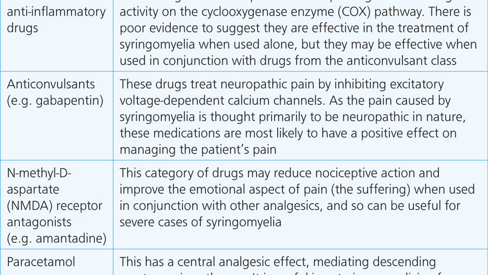
Conservative management generally consists of medical management with multi-modal analgesics, including those prescribed to target neuropathic pain (Rusbridge and Jeffery, 2008), which are often human medications prescribed via the cascade. The medications may be prescribed as part of a multimodal strategy and their role in the treatment of syringomyelia are described in Table 1.


Table 1. Analgesics prescribed for the treatment of syringomyelia
Analgesic class Role in treatment of syringomyelia
Non-steroidal anti-inflammatory drugs These drugs inhibit the production of prostaglandins through activity on the cyclooxygenase enzyme (COX) pathway. There is poor evidence to suggest they are effective in the treatment of syringomyelia when used alone, but they may be effective when used in conjunction with drugs from the anticonvulsant class
Anticonvulsants (e.g. gabapentin) These drugs treat neuropathic pain by inhibiting excitatory voltage-dependent calcium channels. As the pain caused by syringomyelia is thought primarily to be neuropathic in nature, these medications are most likely to have a positive effect on managing the patient's pain
N-methyl-D-aspartate (NMDA) receptor antagonists (e.g. amantadine) This category of drugs may reduce nociceptive action and improve the emotional aspect of pain (the suffering) when used in conjunction with other analgesics, and so can be useful for severe cases of syringomyelia
Paracetamol This has a central analgesic effect, mediating descending serotonergic pathways. It is useful in veterinary medicine for ‘breakthrough’ pain as a ‘rescue analgesic’ to top up existing pain management, and may be useful for acute flare-ups of syringomyelia
(Rusbridge, 2020)

There is anecdotal evidence from owners to suggest that there is a reduction in clinical signs in affected patients prescribed medications that decrease the production of CSF (Rusbridge, 2020). The reasoning behind the prescribing of these medications (examples include furosemide, cimetidine and omeprazole) is that CSF is the driving force behind the pain mechanisms caused as a result of syringomyelia. Further studies in this area are warranted to obtain conclusive evidence.

Owners of patients being managed conservatively are often advised on lifestyle adaptations that may assist with treating the symptoms of syringomyelia. These include:

  • The use of a harness rather than a collar to prevent any pressure from being exerted on the patient's neck which may be painful
  • Raising food and water bowls in order to make access easier for dogs with neck pain
  • Providing non-slip mats or runners on slippery floors to prevent pain flare-ups
  • Ensuring patients are kept at a normal body condition to help prevent additional stress from being placed upon the joints as a result of excessive load
  • Reducing impact and painful flare-ups by ensuring affected individuals do not jump on and off furniture and in and out of the car
  • The avoidance of ‘tuggy games’.

Conservative management may be supplemented with the use of complementary therapies, including acupuncture, and rehabilitation therapies, such as physiotherapy and hydrotherapy (Rusbridge, 2020). The primary aim of adding these treatment techniques is to supplement pain management by addressing secondary soft tissue pain, to help maintain a comfortable range of motion in the neck and to address any neurological dysfunction, such as weakness and proprioceptive deficits.

A study conducted in human patients with syringomyelia revealed that 54% receiving physiotherapy reported an improvement in their symptoms (Smith et al, 2016). This suggests that, although physiotherapy is not effective for all, it is worth considering. A great deal of care must be taken to ensure that syringomyelia patients do not experience an aggravation of pain as a result of any physical therapies. The therapist involved should be qualified, experienced and belong to a professional registration body.

Surgical management

Surgical management of Chiari-like malformation and syringomyelia is generally reserved for patients that have not responded well to conservative management. It may be a viable option for patients experiencing side effects from medications affecting quality of life, patients with pain levels that are not responding to analgesics, or those showing signs of progression of the disease despite conservative management (Hechler and Moore, 2018).

The most common surgical procedure for management of syringomyelia is cranial/cervical decompression (Wolfe and Poma, 2010). The aim is to allow the flow of CSF by removing the supraoccipital bone at the back of the skull, and part of the top of the first vertebrae. It usually has a good initial success rate (figures from studies are quoted at anything from 80–94%) (Hechler and Moore, 2018), but up to 47% of patients appear to have a recurrence of signs related to neuropathic pain within 2 years, thought to be as a result of scar tissue formation (Rusbridge, 2007). It is possible to overcome this through surgical cranioplasty and the use of a titanium mesh to prevent the development of postoperative scar tissue (Wolfe and Poma, 2010).

Prognosis

Syringomyelia is thought to be a progressive condition in the majority of patients, with around 75% of those affected showing signs of progression (Plessas et al, 2012). The majority of patients once on medication are described by owners as having an acceptable quality of life; however, sadly, around 15% of dogs that had been diagnosed with syringomyelia in one study were euthanased as a result of pain (Plessas et al, 2012).

Case study

Signalment

Bert, a 6.5-year-old male CKCS (Figure 1) was referred for rehabilitation therapies in order to manage symptoms, including episodes of yelping when his head was touched and reluctance to climb the stairs. He was receiving medication to control congestive heart failure for which he appeared well managed.

Figure 1. Bert, a 6.5-year-old Cavalier King Charles Spaniel.

Medical history

His medical history from the referring veterinary surgeon detailed a lack of proprioception and neck pain on examination. Bert was prescribed meloxicam and his owner was advised to rest him and bring him back to be examined again in just over a week.

Assessment findings

On initial rehabilitation assessment, Bert was found to have neck pain with minimal available range of motion of the neck. He was particularly reluctant to move his neck towards the left. He repeatedly scratched his neck throughout the course of the assessment visit. He had no evidence of back or joint pain elsewhere, and no neurological deficits present at the time of examination.

When Bert's face shape was analysed, he was considered to have a broad, short skull shape (Figure 2) and a ‘deep stop’, as described above (Figure 3).

Figure 2. Bert had a broad skull shape.
Figure 3. Bert had a pronounced ‘stop’ — a sharp angle between the nasal and maxillary bone and the frontal bone.

Diagnosis

Given his signalment and history, together with assessment findings, syringomyelia was considered the most likely diagnosis for Bert. His owners were not keen to pursue MRI unless Bert's clinical signs could not be resolved, so this diagnosis was not confirmed. Treatment for Bert consisted of:

  • Contacting the referring vet to discuss the prescription of the neuropathic painkiller gabapentin, given the assessment findings
  • Removing Bert's collar and asking his owner to use a harness for exercise purposes (Figure 4)
  • Rehabilitation sessions, which were initially advised at weekly intervals, but reduced in frequency as Bert's condition improved. These included:
  • Gentle range of motion exercises of the neck by encouraging Bert to follow a lure, and asking his owners to continue this at home twice daily to relieve muscle tension in the neck (Figure 5)
  • Massage of the neck muscles and musculature of the forequarters
  • Rhythmic stabilisations, where Bert's weight was pulsed on and off of his forelimbs by the therapist in order to shift his centre of gravity and activate his postural and core muscles, and enhance proprioception. This was performed during rehabilitation sessions, and his owners were asked to continue this exercise at home, twice daily
  • LASER, which was applied to the muscles of the neck to help relieve spasm
  • Bert's owner was asked to restricted his exercise to two to three 20–30-minute walks daily (he had previously been walking for up to 2 hours at any one time)
  • Bert's owner was advised to manage his diet to maintain him at his current weight and prevent him from becoming overweight in the future.
Figure 4. Bert's owners changed from using a collar to a harness.
Figure 5. Gentle range of motion exercises were performed by asking Bert to follow a lure.

Response to treatment

Bert responded to the above treatment extremely well. The number of episodes of yelping per day reduced dramatically, and after 4 weeks the owner reported they had stopped completely. His examination findings at this point revealed a much-improved neck range of motion (although he was still slightly restricted when moving round to the left), and much less tension in the muscles of the neck. Bert's medication, home exercise programme and lifestyle adaptations were continued and his rehabilitation sessions were reduced in frequency to fortnightly, then monthly.

Conclusion

As a result of the potential impact on patient quality of life of syringomyelia, and the outcome of euthanasia if the clinical signs cannot be managed, it is imperative that measures are taken in order to reduce the prevalence of this condition. Increasing public awareness and the use of screening of individuals before breeding appear to be the strategies most likely to be successful in achieving this. The 2008 BBC documentary Pedigree Dogs Exposed had a dramatic effect on increasing awareness of syringomyelia and other conditions affecting pedigree dogs, indicating the positive influence the media can have in this respect.

KEY POINTS

  • Syringomyelia primarily affects the Cavalier King Charles Spaniel, although other toy brachycephalic breeds may also be affected.
  • The main clinical sign is neck pain, which is considered neuropathic in nature.
  • Syringomyelia may be managed conservatively or surgically.
  • Conservative management consists of analgesics for the management of neuropathic pain, drugs that reduce the production of cerebrospinal fluid; and complementary therapies and lifestyle adaptations to help manage the associated symptoms.
  • The condition is progressive, and results in euthanasia in around 15% of cases as a result of pain that cannot be managed.