Case history
The patient was presented to the hospital for routine examination with a history of congestive heart failure (CHF) and advanced tracheal collapse. The owner reported recent exacerbation of the patient's chronic cough; the cough was described as intermittent and dry. Physical examination confirmed a systolic II/VI grade heart murmur, identified severe periodontal disease and an irritable trachea. The veterinary surgeon suspected that periodontal disease was a contributing factor in the exacerbation of the patient's chronic cough and development of tracheobronchitis, as a consequence of bacterial transfer from the oral cavity (Pardali et al, 2010). The patient was admitted to the hospital for dental prophylaxis and extractions under general anaesthesia.
Pre-operative assessment
The patient was receiving pimobendan (Vetmedin, Boehringer Ingelheim Limited) 0.3 mg/kg every 12 hours orally and furosemide (Frusemide, Millpledge Veterinary) 2.5 mg/kg every 12 hours orally for CHF. Pimobendan is an oral inodilator with phosphodiesterase III inhibition, which decreases the breakdown of cyclic adenosine monophosphate (Atkinson, 2009). As a result pimobendan increases cardiac sensitivity to calcium, cardiac contractibility and relaxation, and potentiates arterial and veno-dilatation (Fuentes, 2004), which may exacerbate the hypotensive effects of anaesthetic agents. Furosemide, a loop diuretic, promotes sodium excretion and increases urine output (Smith, 2006), which decreases intravascular volume and preload venous pressure, thereby reducing pulmonary oedema and congestion (Erling and Mazzaferro, 2008). Diuretic use, in conjunction with anaesthetic agents, can promote hypovolaemia, electrolyte imbalance and increase incidence of peri-operative arrhythmia (Groban and Butterworth, 2006).
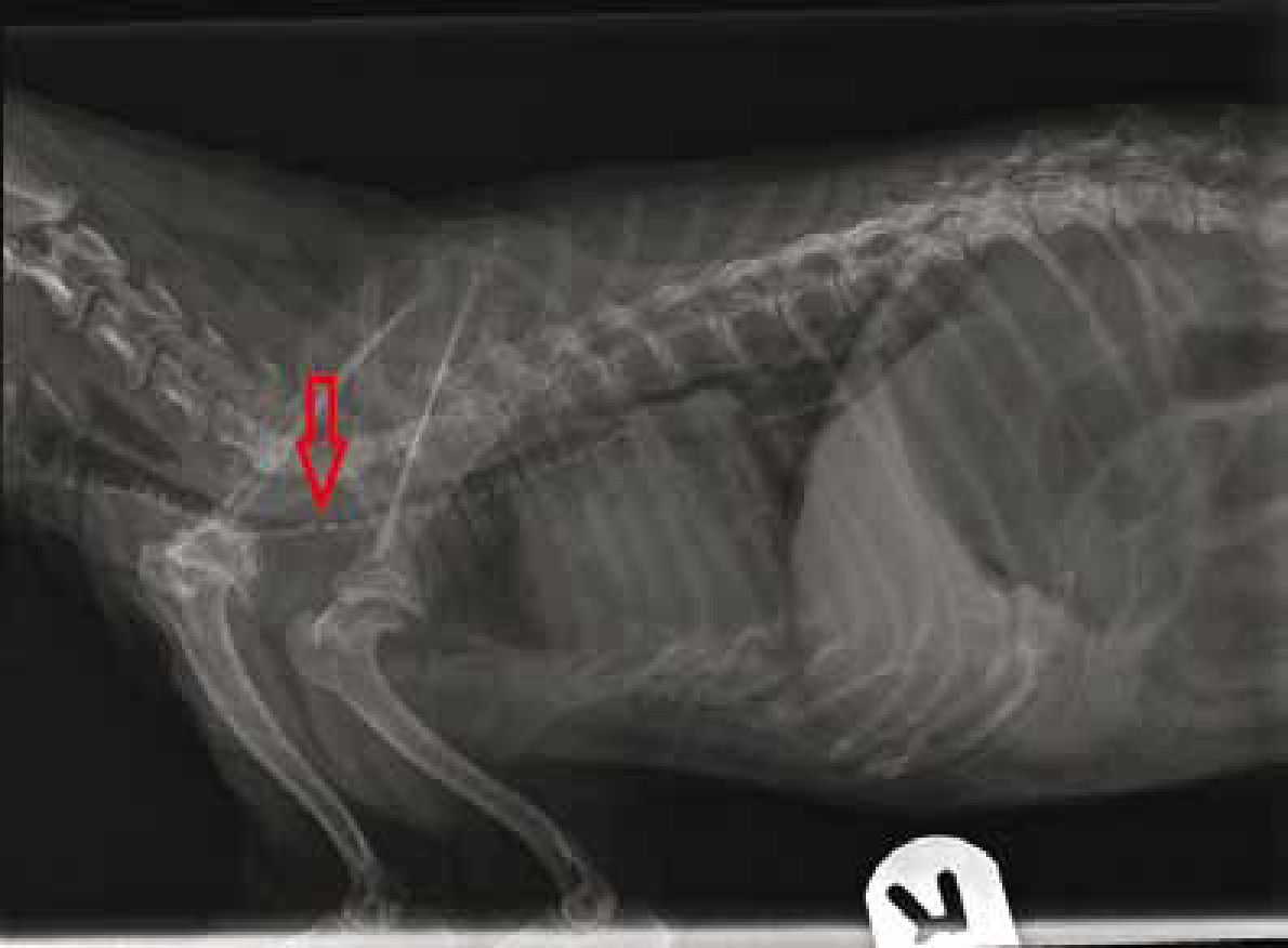
The patient was also receiving diphenoxylate with atropine (Lomotil, Goldshield Pharmaceuticals Ltd) 0.3 mg/kg every 12 hours orally and prednisolone (Prednisolone, Millpledge Veterinary) 0.25 mg/kg every 48 hours orally for concurrent advanced tracheal collapse. Tracheal collapse is characterised by a variable degree of dorsoventral tracheal ring flattening and laxity of the dorsal tracheal membrane (Pardali et al, 2010) (Figure 1). Diphenoxylate is an opioid gastrointestinal motility modifier with antitussive properties, which may reduce damage to the tracheal epithelium generated by chronic irritation (Sun et al, 2008). Administration of diphenoxylate could enhance central nervous and respiratory depression associated with anaesthetic agents (Plumb, 2008), therefore is an important consideration for general anaesthesia in this patient. Atropine, an anticholergenic, can reduce the accumulation of mucus in the lower respiratory tract and act as a bronchodilator (Hardie and Lascelles, 2004). Atropine is included in the diphenoxylate preparation at sub-therapeutic levels to prevent diphenoxylate abuse (Chan, 2008), therefore is unlikely to cause clinically significant cardiopulmonary changes associated with anticholergenic administration. Prednisolone, a corticosteroid, can reduce laryngeal and tracheal inflammation associated with chronic irritation or damage to the tracheal epithelium (Sun, 2008). Non-steroidal anti-inflammatory drugs (NSAIDs) were avoided as concurrent use with corticosteroids can increase the incidence of gastrointestinal ulceration, reduce glomerular filtration rate and prolong clotting times (Narita, 2006).
Laboratory findings indicated elevated alkaline phosphatase (ALP) 180 u/l and blood urea nitrogen (BUN) 39 mmol/l. Elevation of ALP may be attributed to reduced cardiac output or passive congestion of the liver (Goutal et al, 2010) associated with CHF or corticosteroid-induced ALP, which increases serum activity of ALP without causing hepatic dysfunction (Fernandez and Kidney, 2007). In the absence of clinical dehydration, raised BUN value may be indicative of pre-renal a otaemia secondary to CHF attributed to reduced cardiac output or diuretic therapy (DeFrancesco, 2008).
Pre-anaesthetic management
Peri-operative fluid therapy is beneficial to maintain normovolaemia, cardiac output and tissue perfusion, thus counteract the hypotensive effects of anaesthetic agents (Kudnig and Mama, 2002), diuretic-induced hypovolaemia and electrolyte imbalance. Isotonic crystalloid fluid administration is recommended peri-operatively at a reduced rate of 3-6 ml/kg/hour (Kudnig and Mama, 2003) to maintain intravascular volume, electrolyte balance and prevent volume overload. A 24 g intravenous catheter (Jelco, Smiths Medical) was placed in the right cephalic vein to provide patent venous access. Intravenous fluid therapy was initiated (compound sodium lactate (Aquapharm Noil, Animalcare Ltd)) at 5 ml/kg/hour during the peri-operative period, reducing to maintenance rate of 2 ml/kg/hour (Humm et al, 2008) during the postoperative period and continued until self-alimentation was achieved. Fluid therapy was administered using a fluid pump to ensure accurate administration at a pre-determined rate and to prevent volume overload.
Premedication
A low dose of aceproma zine (Calmivet, Vetoquinol) 0.0125 mg/kg and buprenorphine (Buprecare, Animalcare) 0.02 mg/kg was administered via intramuscular injection into the quadriceps muscle. Acepromazine provides adequate sedation, thus decreases patient anxiety and stress-induced catecholamine release, which can result in tachypnoea, reduced myocardial oxygen perfusion and cardiac arrhythmia (Curtis-Uhle and Waddell, 2010). Tachypnoea can increase the negative pressure within the airway, which can exacerbate tracheal collapse and may potentiate complete upper respiratory obstruction (Grubb, 2010). The antagonist effects of aceproma ine on a-adrenergic receptors can promote peripheral vasodilation with subsequent reduction in arterial blood pressure (Grint et al, 2010), which can exacerbate hypotension with concurrent administration of pimobendan. Monitoring of arterial blood pressure was indicated to enable rapid recognition of hypotension (mean arterial blood pressure below 60 mmHg) therefore prevent inadequate tissue perfusion and potential myocardial ischaemia (Shih, 2010). Administration of buprenorphine, a partial opioid agonist, provides analgesia with minimal cardiovascular or respiratory depression; bradycardia and respiratory depression are often associated with administration of full opioid agonists (Waddell, 2010).
Induction and recovery of anaesthesia are critical phases of anaesthetic management in patients with tracheal collapse. During the maintenance phase the patient is intubated and airway patency is maintained (Grubb, 2010). Incidence of hypoxaemia during periods of apnoea or respiratory depression, which may occur during anaesthesia induction or airway obstruction, can be minimised with pre oxygenation — thus increased arterial oxygenation, decreased myocardial workload (Erling and Mazaaferro, 2008) and increased time to desaturation (McNally et al, 2009). The patient was provided with 100% oxygen, by face mask, for 5 minutes prior to and during induction.
Induction
Preparation for patient intubation is crucial in patients with tracheal collapse to enable rapid facilitation of intubation and airway maintenance. Several endotracheal tubes of varying si es, a laryngoscope, lubricant, tie to secure the endotracheal tube and tracheostomy kit were prepared prior to induction. Endotracheal tubes were checked for condition, patency and cuff inflation and were of appropriate length to bypass the tracheal weakness (Grubb, 2010), but not over long so increasing anatomical dead space. Induction was achieved with propofol (Vetofol, Norbrook) 4 mg/kg via slow intravenous administration to effect. Propofol provides smooth induction of anaesthesia; laryngeal relaxation enabled rapid intubation and airway maintenance. The endotracheal tube was appropriately cuffed, over inflation can induce further mechanical inflammation or damage to the tracheal mucosa (Grubb, 2010).
Peri-anaesthetic management
A Humphrey's ADE mini-lack circuit was used and anaesthesia maintained using 2% isoflurane (Isoba, Schering-Plough Animal Health) and oxygen. Isoflurane can cause hypotension via peripheral vasodilation and dose-related myocardial depression, co-administration of nitrous oxide, and supplementary analgesia allowed reduction in the concentration of volatile agent required and therefore improved cardiac output (Monteiro et al, 2010). Nitrous oxide was discontinued 10 minutes before the end of the procedure and the patient maintained on 100% oxygen thereafter to prevent diffusion hypoxia (Keegan, 2005).
During the anaesthetic period the patient's cardiovascular and respiratory parameters were closely monitored, pulse oximetry and capnography were implemented, which enabled assessment of pulse rate and adequacy of ventilation and oxygenation.
Anaesthesia can impair normal thermoregulatory mechanisms by reducing the temperature threshold required to stimulate reflex vasoconstriction via central depression of the thermoregulatory centre of the hypothalamus, and vasodilation associated with anaesthetic agents (Armstrong et al, 2005). Patients weighing less than 5 kg have an increased risk of hypothermia due to their large surface area to volume ratio (Murison, 2001). Hypothermia during the peri and post-operative period can prolong anaesthetic recovery attributed to reduced hepatic drug metabolism (Pottie et al, 2007) and reduced cardiac output (Murison, 2001). During the dental procedure attempts were made to minimise heat loss through evaporation by minimising the wetting of the patient's fur from the drill and scaler water spray. The patient was placed on a VetBed (Petlife International Ltd) to ensure provision of padded bedding with heat retaining properties and enable any in-contact fluid to wick away from the patient. The patient was wrapped in a foil blanket to retain body heat and provide a water resistant barrier. A thermostatically controlled warm air device (Bair Hugger, Advanced Anaesthesia Specialists UK Ltd) was placed over the patient to provide active surface warming and minimise any further heat loss. The patient's core body temperature was monitored during the dental procedure using an indwelling rectal probe.
Caution was exerted with movement and manipulation of the patient during the dental procedure. The patient was disconnected from the anaesthetic circuit when moved or repositioned, to minimise risk of tracheal mucosal damage from cuff rotation (Grubb, 2010). Care was taken not to apply inadvertent pressure on to the patient's thorax, during dental extractions, which could impair the patient's ventilatory efforts.
Advanced periodontitis necessitated extraction of most incisor and premolar teeth; the lower right canine was extracted due to a tooth root abscess which potentiated instability of mandibular symphysis with resultant fracture, which required stabilisation. Analgesia was maintained with buprenorphine 0.02 mg/kg every 6–8 hours via was intramuscular injection. As previously described NSAIDs were avoided due to concurrent corticosteroid use. Due to the patient's small si e some alternative analgesic agents were avoided; such as oral tramadol and transdermal fentanyl, as accurate dosing would have been difficult to achieve. The use of local or regional analgesia could be incorporated as a future intervention to provide effective additional post-operative analgesia (Woodward, 2008). Antibiosis was achieved with cefovecin (Convenia, Pfizer) 8 mg/kg.
Post-anaesthetic management
Isoflurane was discontinued on completion of the dental procedure and 100% oxygen continued; the endotracheal tube remained in situ until the return of the gag reflex, thus indicative that the patient was able to maintain airway patency. Supplementary oxygen was provided to increase inspired oxygen concentration and reduce incidence of hypoxaemia. Pulse oximetry was continued during recovery to monitor for desaturation, which may have indicated tracheal obstruction, and equipment was prepared in case reintubation was necessary.
Requirements for analgesia were assessed at regular intervals, including assessment of the patient's vital signs, gentle palpation of the fracture site, appetite and demeanour, and requirements for analgesia addressed appropriately. The author suggests implementation of a pain scoring model should be considered as a future intervention. Incorporation of a pain scoring model such as the Glasgow composite pain scale (GCPS) would enable standardised and objective assessment of the patient with rapid recognition of pain and intervention as appropriate (Orskov, 2010). Environmental stressors were minimised to reduce pain, anxiety and decrease stress-induced catecholamine release (which as previously described, can induce tachypnoea, reduce myocardial oxygen perfusion and promote cardiac arrhythmia). Of particular consequence is tachypnoea, which can increase the negative pressure within the airway, promote tracheal collapse and induce complete upper respiratory obstruction.
The patient was offered small quantities of a highly digestible low fat diet (Canine Prescription Diet i/d, Hill's Pet Nutrition, Inc.) on recovery. A wet formulation was selected as recommended by Johnson and Hulse (2002) to minimise stress on the healing fracture site. The fracture site was checked after the patient had eaten, and bathed as appropriate with dilute chlorhexidine gluconate (Hibiscrub, Prestige Medical) to remove any food debris. Recovery from anaesthesia was uneventful; the patient was discharged to the owner 24 hours after the dental procedure. The owner was advised to continue a soft or mashed diet and avoid hard foods, chew toys or bones until the fracture site had healed, approximately 8 weeks (Piermattei et al, 2006).The patient was treated on an outpatient basis to administer analgesia (buprenorphine) to maintain comfort.
Conclusion
The author feels that this case highlights the importance of pharmacokinetics (drug absorption, distribution, metabolism and elimination) and pharmacodynamics (drug-induced physiological effects) in the provision of patient care. In the author's opinion, there is a need for increased awareness of pharmacokinetics and pharmacodynamics within veterinary nursing and its practical implication on nursing interventions and patient care.
Anaesthetic management of a patient undergoing dental extractions with concurrent CHF and advanced tracheal collapse must include consideration and assessment of the patient's clinical condition and concurrent medication to enable an appropriate anaesthetic protocol to be implemented. Caution should be exerted not to exacerbate tracheal collapse and endeavour to maintain normovolaemia, normothermia and cardiac output.