

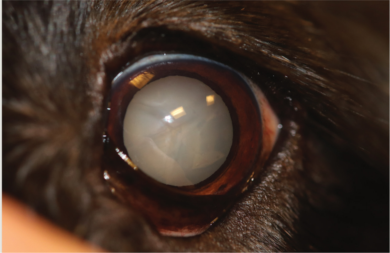
Acataract is a white opacity of the lens which obscures vision by blocking light from reaching the cornea (Moore, 2011) (Figure 1). Cataracts are often hereditary but can also be congenital, developmental, or secondary to metabolic disease (Moore, 2011). Diabetes mellitus is a common cause of cataracts in dogs (Hyde, 2011). An increased glucose concentration in the lens, converts to sorbitol, which acts as an osmotic agent to draw water into the lens, resulting in loss of transparency and cataract formation (Woodham-Davies, 2019). Bilateral cataracts will develop in 50–70% of dogs in the first year following diagnosis of diabetes mellitus regardless of how well the disease is managed (Woodham-Davies, 2019). Cataracts cannot be medically managed, therefore surgical removal of the cataract is indicated (Woodham-Davies, 2019). A number of surgical techniques exist such as extracapsular lens extraction, intracapsular lens extraction and phacoemulsification (Ramani et al, 2013). Case presentation is a deciding factor on which surgical technique is to be used, for example a cataract causing secondary lens luxation to the anterior chamber would likely require an intracapsular lens extraction. Phacoemulsification is the most commonly used method and generally is considered gold standard by veterinary ophthalmologists (Ramani et al, 2013).
Signalment
Species: Canine
Breed: Miniature Dachshund
Age: 12 years 6 months
Sex: Female neutered
Weight: 7.7 kg.
Presentation
On presentation the dog was bright, alert and responsive with a heart rate of 84 beats per minute (normal range 70–140; Crawford, 2011) and respiratory rate of 24 breaths per minute (normal range 10–30; Crawford, 2011). Both eyes were open and comfortable with no discharge present. Menace responses were absent and dazzle reflexes were present in both eyes. Both direct and consensual pupillary light reflexes were present in both eyes. The Schirmer tear test (STT) readings were 18 mm/minute and 19 mm/minute and intraocular pressure (IOP) measured 12 mmHg and 10 mmHg in the left and right eye respectively. A STT reading of 10–15 mm/minute would suggest keratoconjunctivitis sicca (Woodham-Davies, 2020), and IOP measurements >24 mmHg would suggest glaucoma (Sanchez et al, 2017). therefore this patient's values were considered normal. The patient has previously been diagnosed with diabetes mellitus and has since developed bilateral mature cataracts which the owner wished to be removed.
Veterinary interventions
A number of veterinary interventions were carried out:
- Haematology and biochemistry — unremarkable
- Ocular ultrasound — both lens were swollen and echogenic with concentric echoes and a sand-timer appearance centrally. The diagnosis was bilateral cataracts and mild vitreal degeneration
- Presurgical preparation — an hour before surgery, the administration of tropicamide and flurbiprofen sodium (Ocufen®, Allergan Ltd) drops were applied to the eyes alternately every 10 minutes. These drops dilate the pupil and reduce inflammation respectively. Once anaesthetised, povidone iodine (1:50) was used to clean the surface of the eyes, and 1:10 used for the eyelids. The eyes were then irrigated with saline to remove excess povidone iodine. No hair clipping was necessary
- Electroretinograph (ERG) — the ERG was performed under general anaesthetic before the phacoemulsification started. A normal ERG trace was produced, which suggested the patient had a functioning retina and therefore has a good prognosis for vision postoperatively. An abnormal ERG could suggest progressive retinal atrophy (PRA) in which case the patient would be unlikely to be able to see postoperatively, and surgery should be reconsidered.
Nursing considerations
Pain management
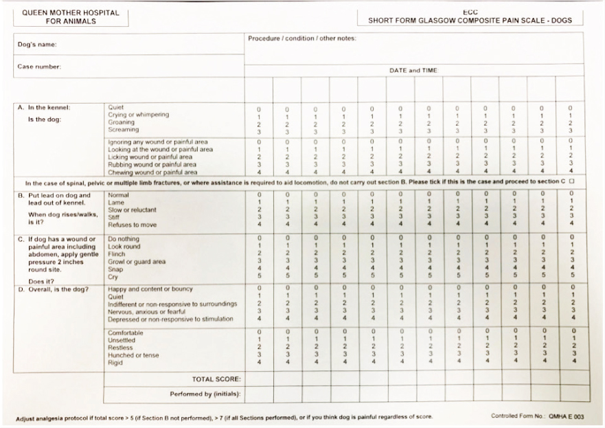
It is well accepted that corneal wounds and glaucoma are painful (Clark et al, 2011; Jacobs, 2017). During phacoemulsification, the cornea is incised in two separate areas so that surgical instruments may access the lens capsule (Woodham-Davies, 2019). Viscoelastic is inserted to inflate the eye (Woodham-Davies, 2019), which can be left behind postoperatively, clogging the drainage angle, and resulting in temporary high pressure. Pain scoring in dogs is often successfully monitored with use of the Short Form Glasgow Composite Pain Scale (GCPS) (Figure 2), and has been reported for use in monitoring ocular pain (Thomas, 2012). A subjective pain score system, created by Clark et al (2011), which is a modified form of the University of Melbourne pain scale, can be used to assess ocular pain in dogs (Figure 3). The pain scale shown in Figure 3 appears more clinically relevant to ophthalmology patients than the GCPS because of its focus on changing ocular-related behaviours.
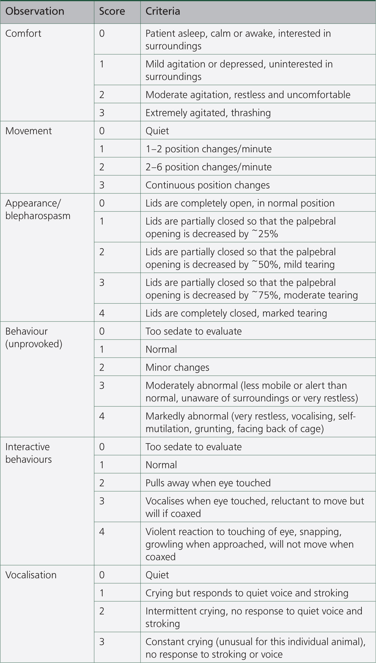
Using the ocular pain score (Figure 3), the patient was considered to be comfortable immediately post-surgery scoring 5/21. The patient demonstrated some mild squinting, however, this could be a result of light sensitivity (Woodham-Davies, 2019). When the kennel door was partially covered with a blanket, the patient fully opened both eyes, suggesting light sensitivity was present. Phacoemulsification results in the release of inflammatory mediators causing inflammation, particularly in diabetic patients (Ganugula et al, 2020). Severe intraocular inflammation was present in the patient's right eye, so a systemic non-steroidal anti-inflammatory drug (NSAID) was administered as these are thought to control ocular inflammation (Clark et al, 2011). Pred forte® (Allergan Ltd), a topical prednisolone, was also prescribed four times daily, however in the author's experience, can sting on application and caused the patient to attempt to rub their eyes following administration. Patients should not be pain scored immediately following topical steroid application as this could give a false high score. The patient wore a buster collar at all times to prevent self-inflicted damage. It should be noted that in human patients, topical steroids have been shown to increase the blood glucose in diabetic patients, therefore, increased monitoring may be required (Bahar et al, 2011).
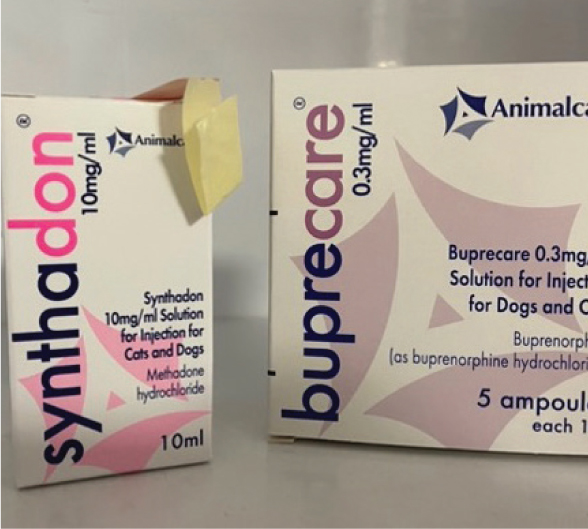
Clark et al (2011) recommended rescue analgesia if the patient pain scores over nine in total, or if a three is scored in any category. Earlier that evening the patient's pain score increased to 9/21 which would indicate the need for opioids (Figure 4). At this time, the IOP of the right eye was 35 mmHg, which was not high enough to trigger the pain relief protocol already set out by the veterinary surgeon. Once the IOP had exceeded 40 mmHg, the registered veterinary nurse (RVN) was permitted to administer buprenorphine at 0.02 mg/kg intravenously. Buprenorphine is a partial mu receptor agonist, which is less suitable than a full mu receptor agonist for management of severe pain in dogs (Clark, 2014). At its peak, the right eye had an IOP of 75 mmHg, and the patient's pain score was 14/21. An IOP of 75 mmHg is an ocular emergency and could result in permanent blindness if not rectified quickly (Woodham-Davies, 2019). If methadone, a full mu opioid, had been given instead of buprenorphine the patient's pain could have been more controlled, however, the noxious stimulus needs to be removed for true pain management (Clark, 2014). Tramadol could also be considered, however, some authors suggest tramadol requires further research in the treatment of ocular pain (Clark et al, 2011).
The veterinary surgeon performed an aqueocentesis on the right eye under sedation, reducing IOP to 7 mmHg. Currently rescue opioids are advised by the veterinary surgeon once IOP reaches a figure of their choosing. In the same surgical ward of a referral hospital, soft tissue and orthopaedic patients are pain scored using the GCPS and analgesia is given in relation to this. For future practice, rescue analgesia should be provided based on ocular pain scoring rather than IOP measurement, as different patients will perceive pain from noxious stimuli differently.
Superficial ocular pain should also be considered in patients following phacoemulsification. The use of the tonometer to measure IOP is not considered painful, however repeated use can cause superficial corneal ulcers, which are considered painful (Clark et al, 2011). A pinpoint ulcer had formed on the patient's right eye, the cause of which is likely to be multi-factoral, including the increased number of IOP measurements being taken as a result of the rise in IOP, theatre lights focused on the eyes making them dry, and opioid usage, which reduces tear production (Woodham-Davies, 2020). Dry eyes are more susceptible to corneal ulcers, meaning this could also have contributed to the creation of a pinpoint ulcer (Woodham-Davies, 2020). Ordinarily topical steroids would not be applied to an ulcerated cornea, however, the Pred forte was continued in order to reduce the intraocular inflammation that was present. Additional analgesics were not necessary, however, the RVN applied lubrication to both eyes regularly to reduce discomfort.
Opioids produce side effects such as nausea, which can lead to inappetence (Hay Kraus and Cazlan, 2019). The diabetic patient should be monitored to ensure pain relief dosages do not impact appetite and subsequently blood glucose levels.
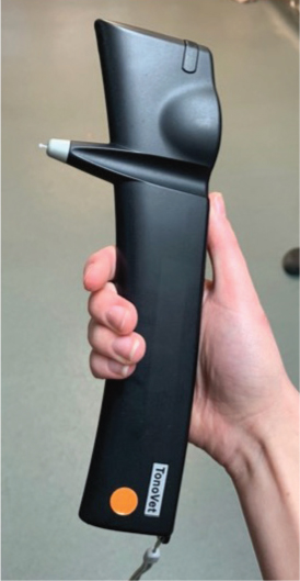
Monitoring intraocular pressure
A rebound tonometer (Figure 5) is used most often in veterinary patients as they are portable, have a small probe and are tolerated well without topical anaesthesia (de Olivera et al, 2018). RVNs generally gain confidence operating the tonometer with regular use, however accurate readings can be difficult to achieve in some patients. At this hospital, a post-surgical IOP curve includes bilateral measurements once every 4 hours over a 24 hour period. This is usually tolerated well by patients as there is sufficient ‘recovery time’ before the next reading. The patient in this report was having IOP measurements taken hourly because of the rise in pressure, between the times of 2100 and 0000, a time when the patient would usually be sleeping. The patient was visibly stressed likely to do with the regular interventions from the RVN, pain associated with high IOP and the unsociable hour.
When restraining ophthalmology patients, minimal contact is desired to reduce risk of damaging delicate ocular structures or increasing IOP (Thomas, 2012). An increased IOP measurement could be caused by restraint around the neck, a neck collar which is too tight or too much pressure on the eyelids. Usually an IOP measurement can be taken by a single RVN but this patient required two RVNs to restrain and measure, in order to use the tonometer at the correct angle. IOP measurements have been shown to be overestimated or underestimated if the reading is taken anywhere other than the central cornea (de Olivera et al, 2018). The RVN measuring the IOP was aware that the reading could be inaccurate, however, knowledge of the surgical procedure (viscoelastic blocking the drainage angle), meant the RVN was confident in reporting a true high IOP.
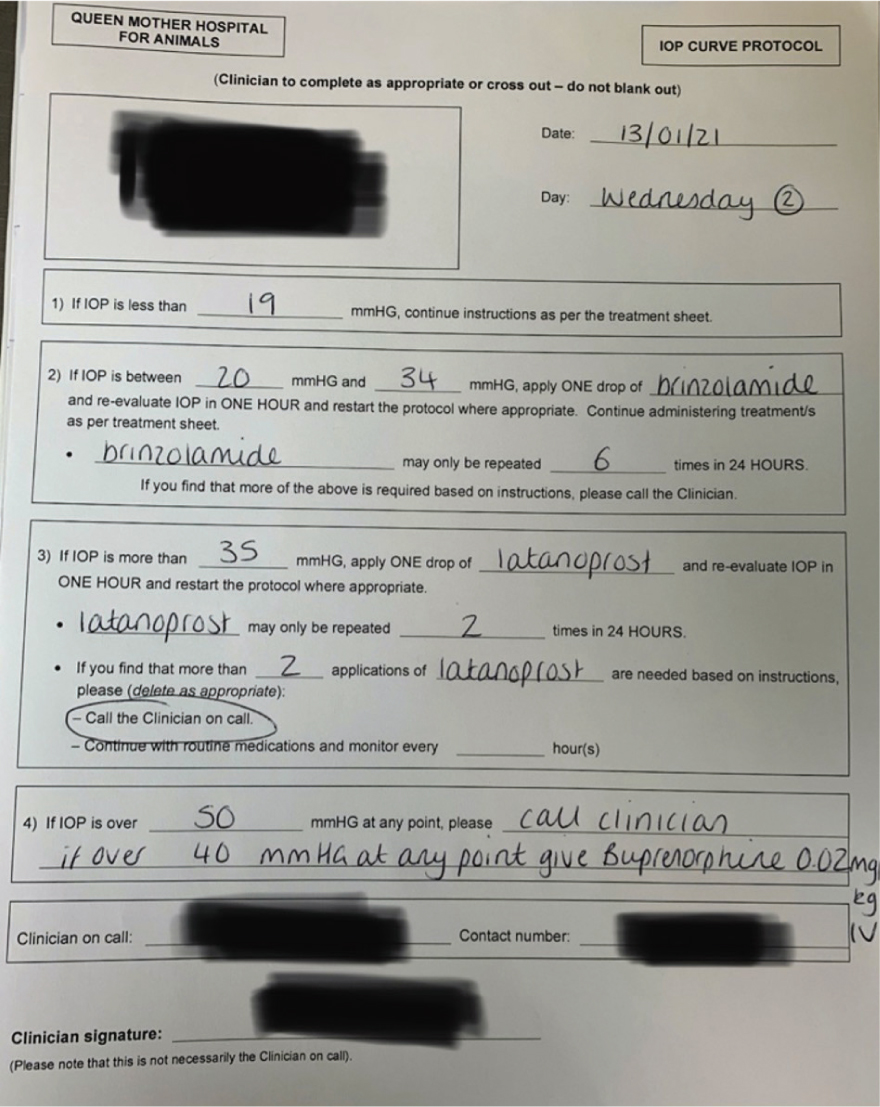
Patients that require IOP measurements will have a protocol written by the veterinary surgeon that instructs RVNs on the appropriate action to take, based on the pressure reading (Figure 6). When the pressure reaches a certain figure the veterinary surgeon will advise additional topical medications, more frequent measurements and rescue opioids if necessary. The patient's IOP was nonresponsive to the additional topical medications and continued to rise to the critical level of 75 mmHg. An elevated IOP in dogs is thought to be over 24 mmHg (Sanchez et al, 2017), however the veterinary surgeon will tailor the values on the protocol to individual patients.
The veterinary surgeon decided to perform an aqueocentesis which would aspirate aqueous humor from the anterior chamber with the purpose of rapidly reducing the IOP (Linn-Pearl et al, 2015). The patient must remain still while the needle is inserted to avoid damage to ocular structures (Linn-Pearl et al, 2015), therefore sedation and topical anaesthesia were required. During this procedure, the role of the RVN was to maintain the patient in lateral recumbency with the affected eye facing upwards, hold the patient's head so that there could be no sudden movements and steady the lid speculum to stop it from falling out of place. Sedation with 0.01 mg/kg of medetomidine intravenously worked well. Butorphanol or methadone could not have been given because of the recent administration of buprenorphine, however with the immediate reduction in IOP, additional analgesics were not required. Aqueocentesis resulted in the patient's IOP reducing from 75 mmHg to 7 mmHg. After the procedure the patient was more comfortable, which contributed to the RVN acquiring IOP measurements with higher accuracy.
Conclusion
RVNs are not able to prescribe analgesia, however they play an important role in assessing how painful patients are and reporting their findings back to the veterinary surgeon. For those who do not work with ophthalmic patients regularly, ocular pain may be difficult to assess. If an ocular pain score was introduced, appropriate pain relief could be administered sooner, which would increase patient welfare. Accurate measurement of IOP is an important skill for RVNs working with ophthalmic patients. An RVN should recognise when an IOP measurement may not be accurate, and be aware of abnormal readings, which should be reported to the veterinary surgeon. Knowledge of the surgical procedure will help RVNs to consider postoperative complications and how they can be managed.
KEY POINTS
- Knowledge of ophthalmic procedures can help registered veterinary nurses tailor nursing care to an individual patient.
- Monitoring ocular pain with a specific ocular pain score could allow better pain management for patients.
- Correct restraint of ophthalmology patients will reduce the risk of an abnormally high intraocular pressure (IOP) reading.
- Lubricant should always be administered following an IOP measurement to reduce the risk of a corneal ulcer forming.