

Nutrition has been recognised as the fifth vital assessment, following temperature, pulse, respiration, and pain, by the World Small Animal Veterinary Association (Freeman et al, 2011). While this campaign has focused on raising awareness of the importance of nutrition for canine and feline patients, it helps bring to mind this aspect of care for any species that is brought into the veterinary practice. Exotics practitioners are no stranger to nutritional needs and disease in exotic companion animals: husbandry-related diseases are disturbingly common in exotic pets. Estimates of the prevalence of husbandry-related diseases range depending on the study, but it is generally agreed that inadequate husbandry is a leading cause of illness in captive exotic animal species (Hartmann, 1993; Fawcett, 2011; Jekl et al, 2011; Prebble, 2011; O'Neill et al, 2020). Client education of appropriate husbandry practices is critical for prevention of husbandry-related disease as well as treatment, however the veterinary staff must be well versed in the needs of these animals in order to provide appropriate information.
Herbivores have many unique adaptations that allow them to survive on a diet with a relatively low calorie density, that is higher in fibre and lower in protein compared with the diet of carnivores and omnivores (Grant, 2010) (Figure 1). These adaptations will be explored to better understand the nutritional needs of exotic companion herbivores.
Summary of target species
The selected species considered in this article are monogastric hindgut fermenters: they have a single, non-compartmentalised stomach, and a large caecum that is the site of microbial fermentation (Carpenter et al, 2012). The following taxa represent some of the most common exotic mammals presented to the veterinary practice as companion animals, and include:
- Order Lagomorpha, family Leporidae (rabbits and hares): the domestic rabbit (Oryctolagus cuniculus) is one of the most common small exotic mammal pets in both the UK (Rabbit Welfare Association & Fund, 2022) and USA (Jones, 2022).
- Order Rodentia, suborder Hystricomorpha, infraorder Caviomorpha. Some texts refer to ‘hystricomorph’ rodents, although this parent clade includes many extinct as well as extant taxa. The infraorder Caviomorpha (the ‘new world hystricomorphs’) includes:
- Caviidae (guinea pigs, cavies, maras, capybara) — guinea pigs (Cavia porcellus) are a common representative of this family as a companion mammal
- Octodontidae (degus, rock rats, and relatives) — the common degu (Octodon degus) is perhaps not as common as the other pet species discussed but is nevertheless well represented in the pet trade
- Chinchillidae (chinchillas, viscachas) — the domestic chinchilla, thought to be descended from the long-tailed chinchilla (Chinchilla lanigera), is a common companion mammal.
Digestive physiology is best described in rabbits, so most of this discussion will be lagomorph-centric. Where the mode of digestion is the same in the species to be discussed, we will extrapolate the general mechanics of how the rabbit digestive system functions to other species. Unique adaptations, anatomy, and dietary requirements of the other featured species will be highlighted.
Gastrointestinal tract adaptations to herbivory
Senses and food selection
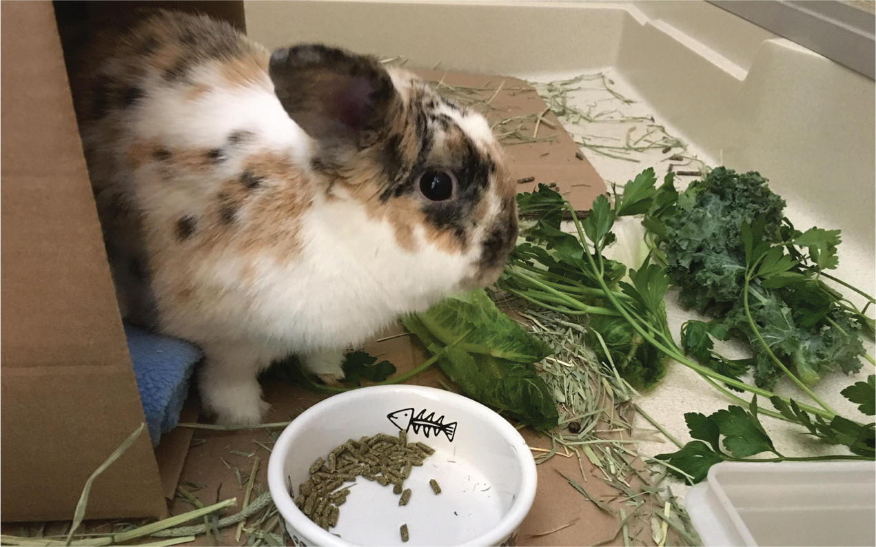
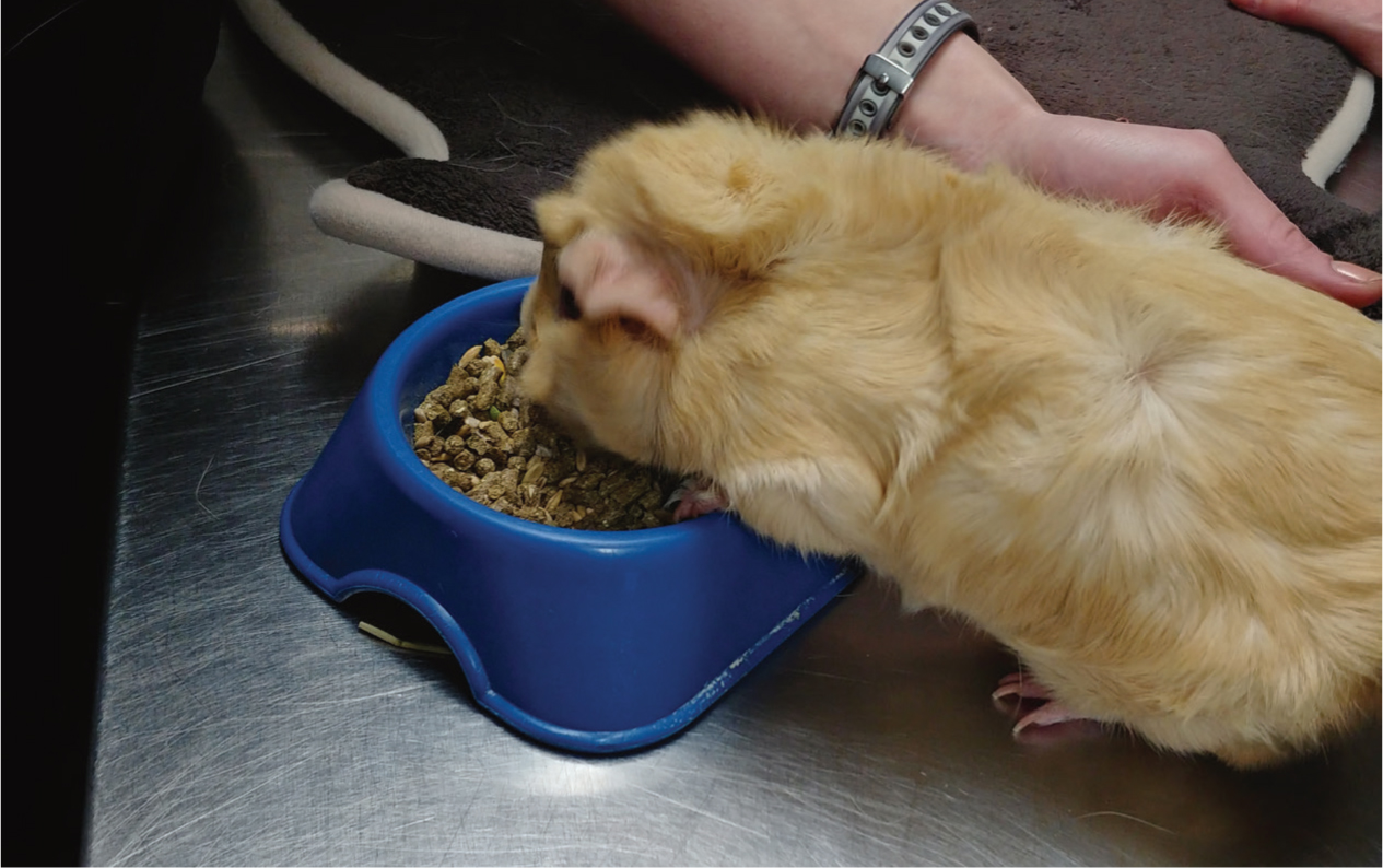
The target species have wide visual fields to facilitate environmental surveillance of threats while foraging, as the low calorie density of the natural diet necessitates the in-take of large volumes of plant material (Carpenter, 2002; Varga, 2014). Rabbits and guinea pigs will use their sensitive lips and vibrissae as well as olfactory cues to distinguish food items, and use their prehensile lips to grasp food. Chinchillas and degus will grasp food with their forelimbs. Rabbits are described as selective feeders and wild rabbits prefer eating tender plants to coarse grasses and sedges (Cheeke, 1987). Selective feeding is also observed in chinchillas where highly palatable food items (e.g. high in simple carbohydrates and fats) are preferred to foods lower in carbohydrates/fats (Wolf et al, 2003). Degus also demonstrate selective feeding behaviour, preferring vegetation that is lower in fibre and higher in protein and moisture (Guitierrez and Bozinovic, 1998). Wild guinea pigs are described as unspecialised grazers (Asher et al, 2004), however in captivity there is vast anecdotal observation that they will preferentially eat concentrates (Carpenter et al, 2012; Quesenberry et al, 2012) (Figure 2).
Rabbits demonstrate a taste preference for glucose and maltose (Laska, 2002). Hunger has several stimuli, including a dry mouth, empty stomach, or decreased blood levels of glucose, amino acids, lactic acid, or volatile fatty acids (Fekete, 1989).
The oral cavity
The vertebrate digestive tract naturally starts at the mouth. Rabbits are known to have a blind spot immediately in front of the mouth (as a result of their wide, lateral field of vision to improve predator detection) and rely on olfaction and sensitive vibrissae to identify food (O'Malley 2005; Harkness et al, 2013; Donnelly and Vella, 2021). Rabbits and guinea pigs have prehensile lips to apprehend food; chinchillas and degus use their forelimbs to apprehend food. These animals feed frequently: rabbits have been described as feeding up to 30 times per day over 4–6 minute intervals (O'Malley, 2005).
The teeth are adapted to a diet that is very abrasive, causing a lot of ‘wear and tear’ to the enamel. Rabbits chew approximately 120 times per minute (Harkness et al, 2013). There are several terms to describe the dentition of these herbivores:
- Hypsodont — the teeth have crowns that extend high past the gingival margin, with short roots (Allaby, 2009)
- Aradicular — the teeth are open-rooted (Harkness et al, 2013).
- Elodont — the teeth grow continuously through the animal's life, to offset enamel wear through their lifetime (Harkness et al, 2013).
The term hypselodont has been used as an all-inclusive term to describe teeth with high crowns, short and open roots, that grow continuously (Janis and Fortelius, 1988) as a result of a germinative bud at the apex containing dental stem cells (Jernvall and Thesleff, 2012; Ungar, 2015).
The incisors are long and used to ‘snip’ plant material, canines are absent, and they possess premolars and molars (that are anatomically indistinct) for grinding food. There is a large gap between the incisors and premolars (the diastema). The tooth is divided into the clinical crown and reserve crown, above and below the gingival margin, respectively (Capello et al, 2005).
Dental formulae of these species can be found in Table 1. Note that rodents are monophyodont and rabbits (Lagomorpha) are diphyodont (Capello et al, 2005).
Table 1. Dental formulae of the featured species
| Species | Incisors | Canines | Premolars | Molars | Total teeth |
|---|---|---|---|---|---|
| Chinchilla, Chinchilla lanigera | 1/1 | 0/0 | 1/1 | 3/3 | 20 |
| Degu, Octodon degus | 1/1 | 0/0 | 1/1 | 3/3 | 20 |
| Guinea pig, Cavia porcellus | 1/1 | 0/0 | 1/1 | 3/3 | 20 |
| Rabbit, Oryctolagus cuniculus, permanent teeth | 2/1 | 0/0 | 3/2 | 3/3 | 28 |
| Rabbit, Oryctolagus cuniculus, deciduous teeth | 2/1 | 0/0 | 3/2 | 0/0 | 16 |
The opening of the mouth in these species is very small, and the lips fold into the oral cavity behind the incisor at the diastema (the inflexa pellita) (Ade, 1999). In rabbits, saliva is continuously secreted by the mandibular salivary gland. All salivary glands (parotid, mandibular, sublingual, zygomatic) produce additional saliva in response to food intake (Davies and Davies, 2003). Rabbit saliva contains amylase that initiates chemical digestion of carbohydrates (Fekete, 1989).
Oesophagus and stomach
The stomach of these animals always contains ingesta (Davies and Davies, 2003), that should have a soft, pliable consistency when palpated. Stomachs that are bloated and turgid, or the contents feel firm and not pliable, are abnormal (Fraser and Girling, 2009; Donnelly and Vella, 2021). Hindgut fermenters have a very tight oesophageal (cardiac) sphincter and cannot vomit or eructate (Davies and Davies, 2003; Donnelly and Vella, 2021). Compared with other non-herbivorous species, they produce a relatively large volume of saliva and stomach fluids that mix with ingesta. These two factors lead to a rapid development of gastric dilatation (of fluids or gas) in the event of digestive hypomotility or obstruction (Kelleher, 2010; Oglesbee, 2010). Guinea pigs are especially predisposed to developing gastric dilatation volvulus (Dudley and Boivin, 2011), although the reason for this is unclear.
Gastric glands produce pepsin and hydrochloric acid. The gastric pH is very acidic: rabbits are reported at <2.0 (Carpenter et al, 2012) and as low as 1.6 (Hamid et al, 2011); Guinea pigs are reported to have a gastric pH of 2.9-4.4 (Merchant et al, 2011). This pH is thought to sterilise ingesta before it reaches the small intestine. When caecotrophs are ingested, the pH rises to 3. Neonatal rabbits' gastric pH is higher at 5–6.5, permitting bacteria to pass through the stomach and small intestine to colonise the caecum (Blas and Gidenne, 2010). Gastric transit time in rabbits is 3–6 hours (Varga, 2014).
Small and large intestines
The small intestines are composed of (cranially to caudally) the duodenum, jejunum, and ileum as in other vertebrate species, and their functions are similar: bile, digestive enzymes, and buffers are secreted here. The duodenum is a site of further digestion of chyme that exits the stomach via the pyloric sphincter. The bile duct empties into the proximal duodenum, and a pancreatic duct opens into the duodenum at the junction of the transverse and ascending loops (Varga, 2014). Motilin is a polypeptide hormone produced by enterochromaffin cells in the duodenum and jejunum, and regulates motility by stimulating smooth muscle contractions of the stomach, small intestine, colon, and rectum in rabbits. Motilin production is stimulated by the presence of fat in the digestive tract, and inhibited by carbohydrates (Adachi et al, 1981; Ito, 1990; Varga, 2014). Lagomorphs possess the sacculus rotundus (or ileocaecal tonsil), a thin-walled, honeycomb-like structure containing lymphatic tissue located at the terminal ileum (Carpenter et al, 2012).
Digesta that moves through the ileocaecocolic junction into the proximal colon is then sorted via a colonic separation mechanism: peristaltic action moves large fibre particles, lignin, and hair further into the colon to become hard faeces, and an antiperistaltic action moves soluble fibres, fluid, and bacteria into the caecum (Quesenbery et al, 2012; Harkness et al, 2013). Lagomorphs utilise a stream of water as a colonic separation mechanism to transport the digesta and bacteria from the proximal colon into the caecum. In contrast, guinea pigs and chinchillas employ a ‘mucus trap’ to recuperate caecal bacteria from the colon (Holtenius and Bjornhag, 1985; Hagen et al, 2016). This is considered less efficient at recapturing bacteria than the lagomorph's mechanism (Franz et al, 2011). The mucus is guided along a colonic furrow in the ascending colon to the caecum (Holtenius and Bjornhag, 1985; Hagen et al, 2016). Hard faeces will pass through the colon, and water reabsorption occurs here as in other vertebrates (Davies and Davies 2003).
The caecum, and caecotroph production
The caecum is the site of hindgut fermentation. Caecal flora possess enzymes (various hydrolases) that break down plant fibres and cell walls via hydrolysis. The resulting monosaccharides and amino acids are used by these flora for energy (Combes et al, 2013). Caecotrophs (sometimes spelled caecotropes, and also referred to as ‘soft faeces’ or ‘night faeces’) are the specialised product of caecal microbial fermentation. Caecotrophs contain sequestered products of microbial fermentation, including volatile fatty acids (VFAs), amino acids, B vitamins, vitamin C, and vitamin K (Varga, 2014; Mayer, 2021). VFAs are absorbed actively through the walls of the caecum and colon and are used for energy (Davies and Davies, 2003).
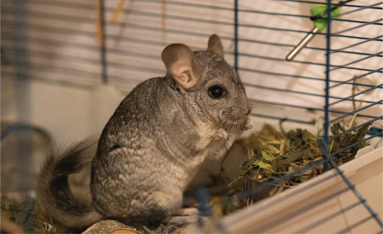
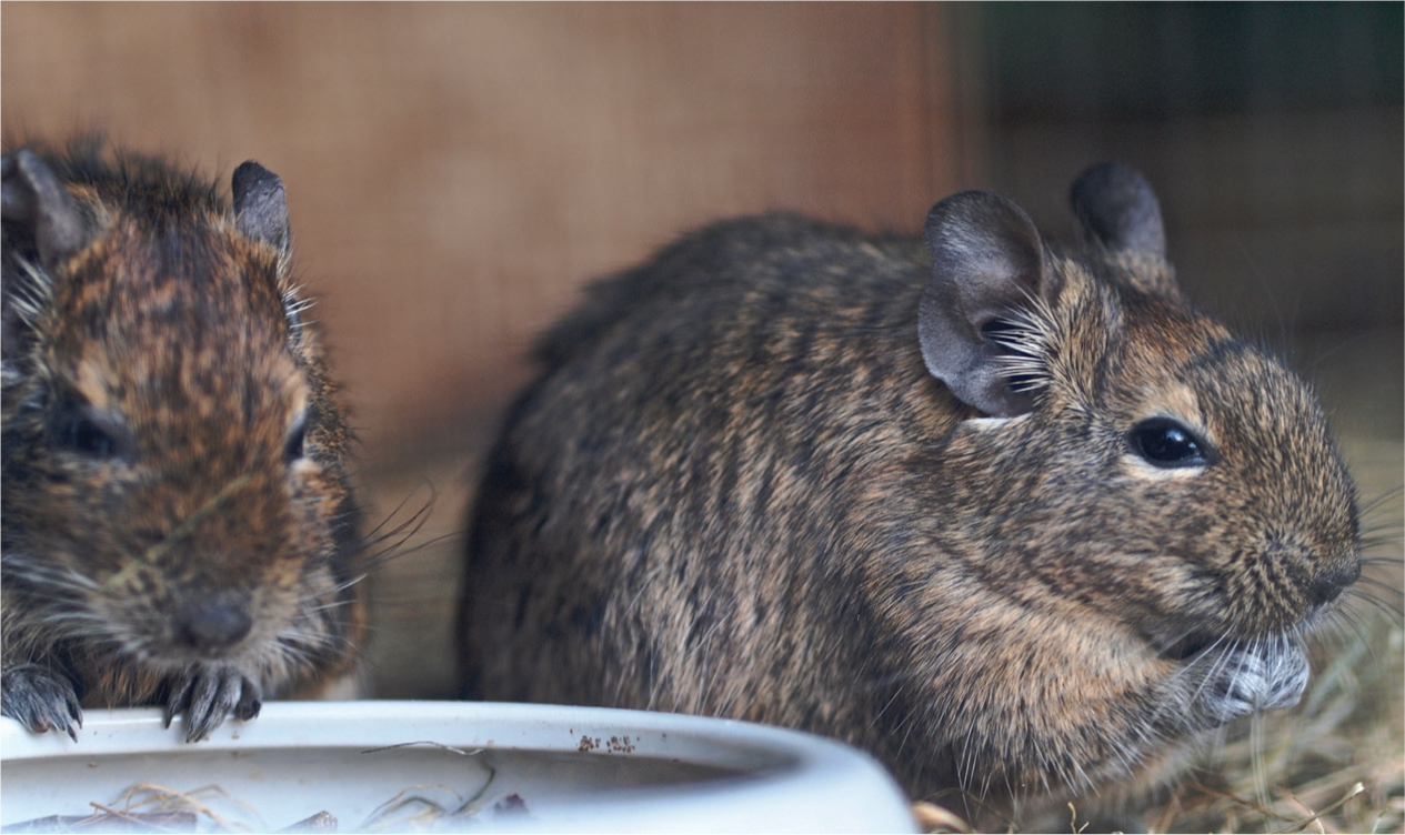
Caecotrophs in rabbits are produced about 4–8 hours after eating (Varga, 2014), and are expelled typically at night and early morning (Davies and Davies, 2003). Guinea pigs are described as eating food and their caecotrophs throughout the day (Ebino, 1993). In contrast, chinchillas have been described as ingesting more than 70% of their daily feed intake at night (Wolf et al, 2003) and typically produce and ingest caecotrophs at night (Quesenberry et al, 2012) (Figure 3). Degus also predominantly produce and ingest caecotrophs overnight (Kenagy et al, 1999) (Figure 4).
Caecotrophs passing through the colon are coated in mucus, produced by goblet cells, that will protect the contents from the acidic environment of the stomach when ingested (Grant and Specian, 1998). The mucus also contains lysozyme that continues breaking down bacteria, even after expulsion from the digestive tract (Camara and Prieur, 1984). The continued lysis of microbial proteins facilitates nutrient absortion while caecotrophs pass through the small intestines (Fekete, 1989). The lagomorph colon has a unique structure called the fusus coli, located at the caudal transverse colon. This muscular, thickened region acts as a sort of ‘gut pacemaker’ and the peristaltic pattern will differ between hard faeces and caecotroph production. It initiates peristalsis throughout the colon, as well as controls separation of fermentable and indigestible digesta as described above (Ruckesbusch and Fioramonti, 1976). The fusus coli is influenced by multiple regulatory mechanisms: circulating prostaglandins, aldosterone and autonomic nervous system are all implicated (Pairet et al, 1986). In guinea pigs, there is experimental data demonstrating that colonic peristalsis appears to be controlled by the autonomic nervous system (Gribovskaja-Rupp et al, 2014).
Caecotrophs are ingested either directly from the anus or from the environment, and the rabbit's masticatory pattern differs to prevent disruption of the mucoid coating (Cheeke, 1987). Although the term ‘coprophagy’ is some-times used in the literature to describe the behaviour of ingesting caecotrophs, it is perhaps important to distinguish this behaviour as caecotrophy and not coprophagy. Coprophagy implies that a waste product is being ingested, and caecotrophs are not waste (Davies and Davies, 2003) but a nutrient-dense product of specific anatomical and behavioural adaptations that is necessary for survival of these species. Ingested caecotrophs will remain in the gastric fundus for 6–8 hours, where further fermentation occurs inside each caecotroph (Varga, 2014).
Identification of normal caecal flora has traditionally been achieved using culture techniques. Modern advances in molecular biology have allowed for a better understanding of the caecal microbiota. It has been estimated that only 24–40% of rabbit caecal flora has been identified using traditional culture techniques (Bagóné Vántus et al, 2014). See Table 2 for a summary of microbes that have been identified as normal flora in the rabbit caecum. Coliform bacteria such as Escherichia coli can proliferate when there is diarrhoea (Mayer, 2020). Rabbits are known to experience bacterial overgrowth (such as E. coli and Clostridium spp.) when fed a low fibre (<12% dry matter basis) and/or highenergy diet (Kelleher, 2010).
Table 2. Summary of gut flora identified in rabbits
| Species identified | Reference |
|---|---|
| Traditional culture techniques | |
| Bacteroides spp. predominant | Fann and O'Rourke, 2001; Sirotek et al, 2001 |
| Bacteroides spp. predominant (Bacteriodetes); Bifidobacterium spp. (Actinobacteria), Endophorus spp., Streptococcus spp. (Firmicutes), Acuformis spp., Clostridium spp., Peptococcus spp., Peptostreptococcus spp., Fusobacterium spp. | Cheeke, 1987, de Blas and Gidenne 1998 |
| Fecal analysis techniques | |
| Flagellated protozoa (Eutrichomastix spp., Enteromonas spp., Retortamonas spp.), amoeboid protozoa (Entamoeba cuniculi) | Lelkes and Chang, 1987; Owen, 1992 |
| Molecular biology techniques | |
| Actinobacteria, Bacteroidetes, Fibrobacteres, Firmicutes, Proteobacteria, Tenericutes, and unclassified microbes | Crowley et al, 2017 |
Conclusion
The digestive tract of monogastric hindgut fermenters is uniquely adapted to optimise the nutrition obtained from an abrasive diet with a low nutrient density. Understanding the intricacies of this digestive mode is essential when considering nutritional counselling for these species as veterinary patients, as well as when investigating digestive tract dysfunction.
The following review article will explore wellness nutrition for these target species.
KEY POINTS
- Husbandry-related diseases, including nutritional disease, are a common cause of illness in exotic companion animals.
- Companion mammal herbivores are predominantly monogastric hindgut fermenters, whose digestive systems are optimised to obtain nutrition from a low calorie, high fibre diet.
- Hindgut fermenters produce caecotrophs in the caecum, which are a source of essential nutrients that may otherwise not be obtained in the natural diet.