
Veterinary physical rehabilitation is full of benefits for animal patients — whether they are recovering from a postoperative procedure, injury, or going through the natural ageing process. As physical rehabilitation veterinarians and nurses, we set up individual goals for our patients to help them gain a better quality of life. One goal that is often part of the rehabilitation plan is the alleviation of pain (Prydie and Hewitt, 2015). When an animal is no longer in pain, he or she is more likely to relax and perform better during exercise (Sluka and Walsh, 2003).
There are many ways to help alleviate pain during a rehabilitation appointment. Transcutaneous electrical nerve stimulation (TENS) can be used in conjunction with medications and other modalities to help alleviate pain before, during and after exercise, or in phases of healing. For the purpose of this article, a brief overview of low intensity, high-frequency TENS (also called conventional TENS) and its clinical application on canine patients, will be discussed (Johnson, 2007).
Origin of TENS
In human medicine, electricity for pain relief has been around since the mid-1800s. Electrical stimulation for pain was not fully accepted by the medical community until a 1965 publication by Melzack and Wall, which discussed the effect it had in response to the gate theory of pain and how TENS can relieve pain (Sluka and Walsh, 2003). Further publications have since emerged researching the gate control theory and its efficacy. Dickenson (2002) and Mendell (2014) are just two of the many articles that have investigated this theory.
As part of a response to Melzack and Wall's (1965) gate theory, an array of animal studies in veterinary medicine have emerged. These animal studies have been used to investigate parameter manipulation and the neurobiological mechanisms underlying the effects of TENS (Walsh, 2003). Animals have been part of electrotherapy research for roughly as long as humans have. Publications such as Ansari et al (2013) and Cassu et al (2012) show evidence of animals being an important part of electrotherapy research.
How does TENS work?
TENS is one type of electrotherapy used in animal rehabilitation. This technique passes electrical currents across the intact surface of the skin to activate underlying nerves (Johnson, 2007). Using low frequencies of electricity to provide pain relief, this therapy is a non-pharmacological treatment for pain control (Sluka et al, 2013).
The gate control theory of pain further describes the efficacy of TENS (Melzack and Wall, 1965; Wall, 1978). This theory suggests that stimulation of afferent nerve fibres with a large diameter inhibit nociceptive nerve fibreevoked pain responses in the dorsal horn of the spinal cord (Sluka and Walsh, 2003; Goldberg and Tomlinson, 2018). Essentially, TENS stimulates the larger, faster-moving cutaneous nerve fibres, and interrupts the slower transmission of the small cutaneous nerve fibres by which pain signals are carried (Goldberg and Tomlinson, 2018).
Clinical application
When deciding whether a patient is a candidate for TENS, a full physical examination should be performed with special focus paid to pain assessment. An incomplete evaluation of location and intensity of pain may result in failure of treatment (Goldberg and Tomlinson, 2018). Pain should continue to be assessed for the duration of the TENS treatment. The patient's status of pain during treatment will help monitor its effectiveness (Levine and Bockstahler, 2014). Whether the patient is in acute or chronic pain should be considered as this will affect treatment time. It should be taken into consideration that TENS is most helpful when the current is being actively administered. The patient should thus be receiving TENS as an adjunct therapy; the use of this electrotherapy alone for pain relief is not considered physical therapy (Sluka and Walsh, 2003).
TENS should be considered for a patient when pain reduction is needed to improve participation in treatment; in conjunction with pain medications to help with pain wind up; or in patients that do not tolerate pain-relieving medications (Niebaum, 2013). This type of treatment can be useful for pain of nociceptive, neuropathic and musculoskeletal origin (John-son, 2007).
Contraindications and adverse side effects of TENS need to be considered before its use on a patient. Contraindications (Niebaum, 2013) may include:
The application of TENS rarely has any side effects in veterinary patients. A side effect that may develop is skin irritation and contact dermatitis beneath the electrodes (Niebaum, 2013). If this happens, the patient's veterinarian should be consulted and treatment discontinued for the time being.
Electrode placement and machine setup
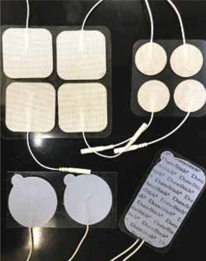
Different types of electrode pads include rubber, gel, and brush-like needle pads. The rubber and gel electrodes require the treatment area hair to be clipped. Once clipped, clean the skin with a little alcohol or water to remove dirt and debris from the treatment area. If the pads do not come with gel, a contact medium, such as ultrasound gel is safe to use and should be applied evenly under the pad to ensure uniform transmission (Levine and Bockstahler, 2014). In circumstances where the animal's hair cannot be clipped, a greater amount of a contact medium may be used to try to achieve adequate conduction. If the needle pads are used, clipping the hair is not necessary. The skin should be wetted with water or alcohol before using these pads (Levine and Bockstahler, 2014). Although needle pads are an acceptable type of electrode to use, they are not usually supplied with the machine and are therefore rarely used by practitioners.
When it comes to the shape and size of the electrode pad, the treatment area needs to be considered. Pads range from very small, round, oval, square, and rectangular to larger pads of the same shapes (Figure 1). Most electrode pads can be easily cut into smaller sizes or different shapes to cover the treatment area. The pads should have low resistance; be flexible enough to conform to the tissue; and be highly conductive and reusable (Goldberg and Tomlinson, 2018).
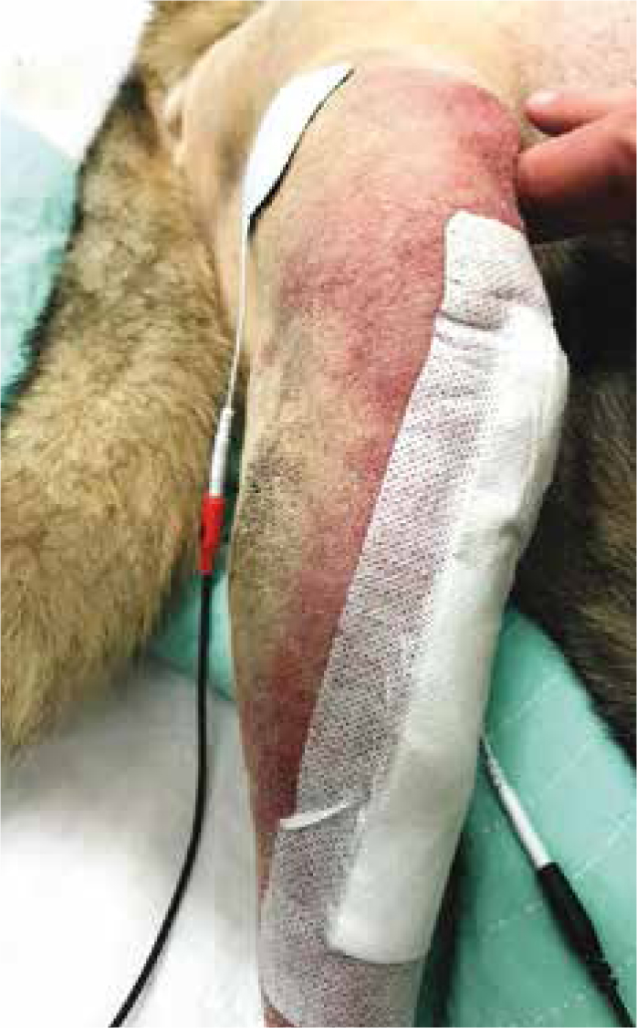
Placing the pads in the proper location is key to an effective outcome. The electrode pads may be placed on relevant dermatomes, along the main nerves proximal to the sight of pain, paravertebrally, or to contralateral ‘mirror’ sites (Johnson, 2007). For acute pain, consider placing the electrode pads parallel to the incision or right over the source of pain, but not on or directly over a wound. In patients with chronic pain, consider placing the pads directly over or proximal to the sight of pain. It may be necessary to try several locations before achieving the desired effect (Levine and Bockstahler, 2014).
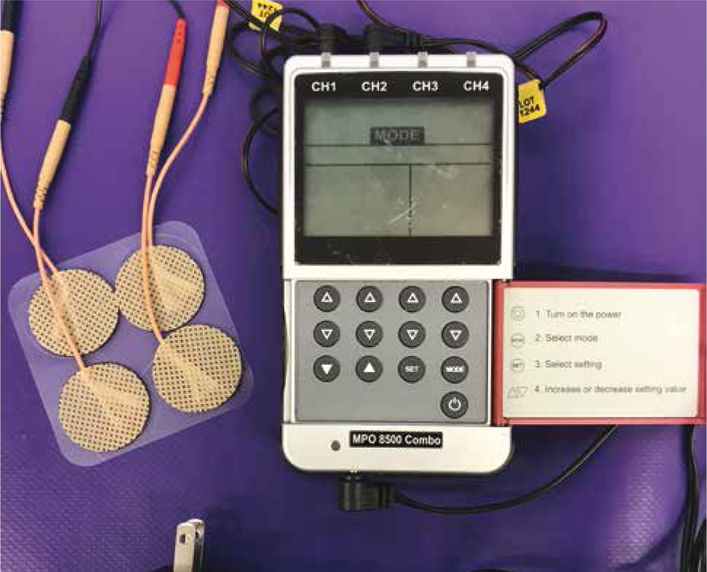
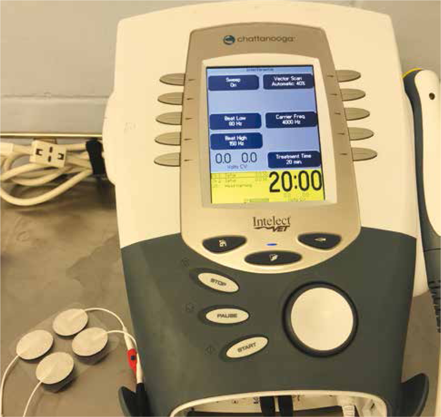
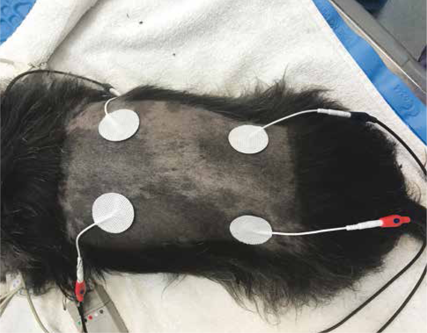
There are two electrode pads that attach to each channel on the machine. Most TENS machines include one black and one red electrode per channel. If a machine does not come with coloured electrodes, they may still be used; the position of the red and black electrode has little effect in clinical practice as biphasic currents are used (Johnson, 2007) (Figure 2; Figure 3). Common biphasic currents used in veterinary medicine include the use of one channel with two electrodes or two channels with four electrodes. The decision of how many electrodes are needed for a treatment can be determined by the location and size of the treatment area, as well as the size of electrode pads. Two electrodes are usually reserved for smaller areas, i.e. smaller muscle bellies and joints (Figure 4); whereas four electrodes are reserved for larger areas, i.e. back, neck, and other large muscle bellies (Figure 5).
When using two electrodes, the pads are placed directly parallel to each other. If four electrodes are used, the two electrodes from the same channel are either placed parallel to each other or in an ‘X’ pattern; thus crossing the channels gives maximum effect in the centre of the large treatment area. Both two and four electrodes may be used to apply a type of conventional TENS to a patient (Levine and Bockstahler, 2014).
Once the electrode pads are placed on the treatment area and the proper current is chosen, the settings or the treatment can be determined. Manipulating the frequency, pulse duration, and amplitude will determine what kind of TENS will be initiated (Goldberg and Tomlinson, 2018). Commonly used conventional TENS settings include stimulation parameters that are low-intensity, high-frequency, and short-pulse duration (Walsh, 2003). These parameters should create a comfortable sensation with no muscle contraction and should use an amplitude just high enough to elicit a sensory response (Goldberg and Tomlinson, 2018). In conventional TENS, the large nerve fibres are stimulated using low-current intensities and high frequencies. Therefore, any frequencies greater that 50 Hz will activate these fibres, thus activating the gate control theory (Levine and Bockstahler, 2014). Another form of TENS, that may be used if patients are not responding to conventional TENS, consists of high-current intensities and low frequencies, also referred to as ‘acupuncture TENS’. This type of TENS stimulates the A delta fibres resulting in endogenous release of opioids into the spinal cord (Johnson, 2007; Watson, 2016).
An example of parameter settings for conventional TENS includes a frequency of 30–150 Hz, pulse duration of 50–100 µs, and an amplitude just below the sensory response threshold. Duration of treatment will vary based on how the patient is responding and what kind of activity, if any, is being performed (Niebaum, 2013; Levine and Bockstahler, 2014; Pyrdie and Hewitt, 2015; Goldberg and Tomlinson, 2018). Recommended treatment durations vary as well. In acute cases, TENS may be administered up to two times daily for 15 minutes; in chronic cases, it may be a 15–60 minute daily session (Levine and Bockstahler, 2014). Patient response to treatment will help to evaluate if the correct duration is chosen.
Appropriate machine and electrode maintenance
Basic cleaning and maintenance should be carried out for optimal use of the machine chosen and may include:
Troubleshooting TENS
For the veterinary professional, troubleshooting modalities can be a common job in the hospital. Luckily, there are only few things to troubleshoot in a TENS machine.
If the patient does not seem to be receiving enough pain relief or seems to be uncomfortable during treatment, check the machine settings. Decreasing or increasing intensity can make a big difference to a patient's level of comfort. Make sure the electrodes are in the proper position and in appropriate contact with the skin. A contact medium, such as ultrasound gel, may be needed to increase transmission. If the TENS machine being used is battery-operated, try checking the battery or plugging in the unit where possible.
Conclusion
Electrotherapy has been a part of both human and animal medicine for centuries — proving that it is a helpful modality to use when trying to relieve pain. Research on the efficacy and use of TENS in animals is still ongoing. However, TENS is an effective modality to have available for patients in pain because it is inexpensive, easy to maintain, comfortable for the patient, and has minimal side effects. Having patients that are pain-free increases their quality of life and their ability to perform during rehabilitation.