References

Aaron A, Eggleton K, Power C, Holt PE. Urethral sphincter mechanism incompetence in male dogs: a retrospective analysis of 54 cases. Vet Rec. 1996; 139:(22)542-546 https://doi.org/10.1136/vr.139.22.542

Adams L. Cystoscopy, 3rd edn. In: Elliott J, Grauer GF, Westropp JL (eds). Gloucester, England: BSAVA; 2017

Bartges JW, Callens AJ. Urolithiasis. Vet Clin North Am Small Anim Pract. 2015; 45:(4)747-768 https://doi.org/10.1016/j.cvsm.2015.03.001

Berent A, Mayhew P. Medical and surgical management of urinary incontinence, 3rd edn. In: Elliott J, Grauer GF, Westropp JL (eds). Gloucester, England: BSAVA; 2017

Byron J. Diagnostic approach to the incontinent patient, 3rd edn. In: Elliott J, Grauer GF, Westropp JL (eds). Gloucester, England: BSAVA; 2017

Cannizzo KL, McLoughlin MA, Mattoon JS, Samii VF, Chew DJ, DiBartola SP. Evaluation of transurethral cystoscopy and excretory urography for diagnosis of ectopic ureters in female dogs: 25 cases (1992–2000). J Am Vet Med Assoc. 2003; 223:(4)475-481 https://doi.org/10.2460/javma.2003.223.475

Daniels L. Nursing of the ectopic ureter patient. Veterinary Nursing Journal. 2014; 27:(9)333-356

Davidson AP, Westropp JL. Diagnosis and management of urinary ectopia. Vet Clin North Am Small Anim Pract. 2014; 44:(2)343-353 https://doi.org/10.1016/j.cvsm.2013.11.007

Ettinger SJ, Feldman EC. Textbook of veterinary internal medicine: Diseases of the dog and the cat, 7th edn. United States: Elsevier Health Sciences; 2010

Fischer JR, Lane IF. Urinary incontinence and urine retention, 3rd edn. In: Elliott J, Grauer GF, Westropp JL (eds). Gloucester, England: BSAVA; 2017

Gear R, Mathie H. Medical disorders of dogs and cats and their nursing, 5th ed. In: Turner L, Cooper B, Mullineaux E, Greet T (eds). Gloucester, England: BSAVA; 2011

Gregory SP. Developments in the understanding of the pathophysiology of urethral sphincter mechanism in competence in the bitch. Br Vet J. 1994; 150:(2)135-150 https://doi.org/10.1016/S0007-1935(05)80222-2

Holt P. Urinary incontinence in the dog. In Pract. 1983; 5:(5)162-173 https://doi.org/10.1136/inpract.5.5.162

Hotston Moore A, England G. Rigid endoscopy: urethrocystoscopy and vaginoscopy, 1st ed. In: Lhermette P, Sobel D (eds). Gloucester, England: BSAVA; 2015

Martinez C, Bennaim N, Shiel R. Urinary incontinence in dogs: pathophysiology and medical management. The Veterinary Nurse. 2015; 6:(8)470-480 https://doi.org/10.12968/vetn.2015.6.8.470

McLoughlin MA, Chew DJ. Surgical views: surgical treatment of urethral sphincter mechanism incompetence in dogs. Compend Contin Educ Vet. 2009; 31:(8)360-373

Nelson RW, Couto GC. Small animal Internal Medicine, 4th edn. United States: Elsevier Health Sciences; 2008

Ectopic ureters and ureteroceles in dogs: treatment. 2004. https://www.vetfolio.com/learn/article/ectopic-ureters-and-ureteroceles-in-dogs-presentation-cause-and-diagnosis (accessed 26 January 2023)

Canine urinary incontinence: diagnosis and treatment
02 February 2023
14 mins read
Volume 14 · Issue 1
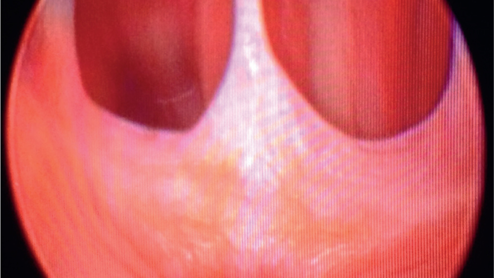
Figure 1. Vaginoscopy reveals a conformation abnormality leading to incontinence; this is a vaginal septum caused by a persistent hymen.

Abstract

Canine urinary incontinence is a common presentation in small animal practice. The care required by the owners at home should not be underestimated as a number of these dogs are presented by owners with a request for euthanasia. Many of the causes of incontinence are treatable, so the veterinarian and veterinary nurse should perform a thorough investigation in order to obtain a diagnosis and instigate appropriate therapy. This article outlines the initial approach to an incontinent dog and discusses the specific diagnostics and treatment options available and nursing care required.

Urinary incontinence is defined as the involuntary passing of urine (Gear and Mathie, 2011). It is a condition commonly encountered in small animal practice, and can be associated with anatomical, physical, inflammatory and neurological disorders (Holt, 1983; Nelson and Couto, 2008). The causes of urinary incontinence are seldom life threatening, but the management of such conditions can be extremely hard work and require a lot of patience from owners. Consequently, a lot of owners request euthanasia. To prevent unnecessary and potentially premature euthanasia of an otherwise relatively healthy dog, it is important to identify the cause and any concurrent issues that may be contributing to the problem and making it worse, so that appropriate treatments may be started, and signs and symptoms eased. This should be done promptly and systematically.

Pathophysiology

To understand how a dog becomes incontinent it is important to first understand the physiology of normal micturition (the act of urinating). Urinary continence requires coordination between the urinary bladder and the urethral sphincter mechanism to allow the passive storage and active voiding of urine (Nelson and Couto, 2008). There are two phases of micturition: the storage phase and the voiding phase.

Storage phase

During the storage phase, urethral tone must exceed the pressure within the bladder for continence to be maintained (Holt, 1983). During this phase the bladder slowly fills up and the urethra stays closed, not allowing urine to pass (Fischer and Lane, 2017). The bladder relaxes while it is filling so there is no urgency to urinate. Mechanical properties, such as the length of the urethra and engorgement of the urethral venous plexus, also contribute to the maintenance of urethral resistance (Holt, 1983). Structural and functional abnormalities during this phase can cause signs of urinary incontinence. Reasons for incontinence in this phase may include:

  • Failure of the urethra to close during storage (urethral sphincter mechanism incompetence)
  • Anatomical abnormalities of:
  • where the ureters terminate
  • bladder shape or size
  • urethral shape and size
  • Urinary bladder dysfunction (Berent and Mayhew, 2017).

Voiding phase

During the voiding phase the bladder receives the message to contract and this is when the need to urinate is felt, resulting in urethral relaxation (Nelson and Couto, 2008) and subsequent urination happening. Urine voiding disorders tend to present as urinary retention (Fischer and Lane, 2017), usually resulting in urinary overflow incontinence. Overflow incontinence is usually a result of either mechanical obstruction, such as urinary calculi or strictures, or neurological disorders, such as dysautonomia and detrusor-sphincter reflex dyssynergia (Brown, 2023).

Investigations

History

A comprehensive history should be taken to allow the veterinarian to differentiate between true incontinence and abnormal urination as a result of polyuria, polydipsia or behavioural reasons. The history will allow the veterinarian to gather clues to a possible diagnosis and that will help direct their choice of investigations.

Questions to ask the owner

  • Signalement (age, breed, sex)
  • Reproductive status
  • Duration of the incontinence – since birth, following neutering, acute onset
  • Nature of the incontinence – frequency, amount passed and when during the day or night the dog is incontinent
  • Urination – normal vs pollakiuria, stranguria, dysuria or haematuria
  • History of trauma
  • Previous urinary tract disease
  • Other clinical signs – neurological deficits, polyuria, polydipsia, tenesmus, vaginal discharge and weight loss.

Clinical examination

A thorough clinical examination is required in all patients. This should include inspection of the external genitalia and surrounding coat for evidence of incontinence such as wetness, skin excoriations and dermatitis. If the incontinence is intermittent no abnormalities may be seen (Holt, 1983).

Where possible, the dog should be observed urinating and the bladder palpated before and after voiding to assess bladder size, tone and position. If a distended bladder is found, it is useful to establish whether the bladder can be expressed. Those that are easily expressed are often associated with decreased detrusor contractility. Alternatively, if the bladder is difficult to express, a functional or physical obstruction may be suspected (Nelson and Couto, 2008).

All male dogs should also have their prostate and urethra palpated via the rectum and females should have a vaginal examination to identify any masses, strictures and conformational abnormalities. Figure 1 shows a congenital abnormality of the vaginal septum called a persistent hymen, allowing urine to pool and retain within the vagina, causing frequent urine infections and incontinence. The body condition score should be considered as incontinence can be exacerbated by obesity (Gregory, 1994). Finally, a neurological examination should be performed if neurogenic incontinence is suspected. This examination should include assessment of anal tone, spinal reflexes and hind limb proprioception (Holt, 1983).

Figure 1. Vaginoscopy reveals a conformation abnormality leading to incontinence; this is a vaginal septum caused by a persistent hymen.

Urinalysis

Urinalysis should be performed in all cases of urinary incontinence (Byron, 2017). As a minimum it should include a urine specific gravity, dipstick and sediment exam and can be carried out by a veterinary nurse. This will provide important information regarding the presence of inflammation, infection, crystals and hydration status. A urine sample for culture and sensitivity should also be obtained as urinary tract infections are frequently seen with urinary incontinence (McLoughlin and Chew, 2009). A voided urine sample may be collected by walking the patient and catching mid stream urine in a sterile kidney dish, then the sample can be placed into a plain and boric urine tube and refrigerated until ready to analyse.

Routine blood work

Serum biochemistry is useful if other causes of abnormal urination are considered, such as renal disease or endocrine disorders. Haematology is often non-specific but can provide information about inflammation. Both haematology and biochemistry may also be useful if the patient has decreased urine concentration or dehydration (Byron, 2017). Veterinary nurses can perform venepuncture; ideally the jugular vein should be used as it is a larger vessel and is generally considered to be better for sampling. Otherwise, any vein that is palpable can be used – the most common sites include the lateral or medial saphenous vein or the cephalic vein.

Radiography

Both plain survey and contrast radiographs of the urinary tract are useful in the investigation of urinary incontinence. Survey radiographs should be taken first to evaluate the bladder, prostate, urethra, pelvis and spine for obvious abnormalities such as a mass or calculi (Ettinger and Feldman, 2010). These can be performed by a veterinary nurse with the patient either under sedation or anaesthesia. The views most commonly used are ventrodorsal and lateral views of the abdomen.

Contrast studies of the urinary tract are performed under general anaesthesia. Options include intravenous urography, urethrocystography or vaginourethography (Ettinger and Feldman, 2010). A pneumocystogram (negative contrast) will allow interpretation of the prostate and bladder position and wall thickness. Positive contrast, on the other hand, will provide information about mucosal detail, highlight filling defects of the urethra and identify ureteral strictures (Figures 2a and b).

Figure 2. A double contrast study – (a) right lateral and (b) ventrodorsal – revealing a markedly intrapelvic bladder. This is more apparent on the ventrodorsal view.

To help with interpretation, the dog should be starved for a minimum of 12 hours and have an enema to ensure the colon is empty (Holt, 1983). Both warm water enemas and commercial enemas such as a Fleet enema are appropriate for this procedure. The patient has the required enema contents placed into the rectum, ideally this procedure should be done outdoors. Multiple enemas may be required and can be carried out in the hospital overnight or first thing in the morning to ensure a clean colon. Once the contents of the colon are clear in colour the enemas can stop. However, if they are not well tolerated by the patient consciously, they can be done once anaesthetised.

Ultrasound

Ultrasound can be useful for evaluating the bladder wall, prostate, regional lymph nodes and identifying calculi or masses (Ettinger and Feldman, 2010). In experienced hands evaluation of the ureters and their point of insertion is possible. Veterinary nurses can assist in restraining these patients for ultrasonography. Depending on temperament some patients may require light sedation – these patients will require monitoring while sedated.

Computed tomography

In better-equipped specialist clinics, this advanced imaging tool can be useful for examining the urinary tract. The veterinarian will be able to see the structures of the abdomen in better detail and at several different angles (Byron, 2017). An intravenous urogram can further aid interpretation of the urinary structures, for example in the identification of ureteral ectopia (Bartges and Callens, 2015). This allows the veterinarian to determine if the urinary incontinence may be fixed surgically or not.

Cystoscopy

Cystoscopy allows direct visualisation of the lower urinary tract (vagina, urethra, bladder and ureteral orifices), it is minimally invasive and performed under general anaesthesia for patient comfort. Adequate pain relief in the premedication should be considered for these cases as these procedures can be quite painful. Opioids such as methadone work quite well for this.

Cystoscopy is technically easier in female dogs because the large endoscope size makes the scope easier to pass. Cystoscopy in female dogs is usually performed with a rigid scope. Urethrocystoscopy can occasionally be performed in larger male dogs if a narrow enough flexible endoscope, such as a bronchoscope, is available. In male dogs the urethra is long, narrow and curved, making this procedure much more difficult (Hotston Moore and England, 2015).

The area around either the vulva or prepuce should be clipped and surgically prepared. The cystoscope and all the equipment required should be sterile so as not to introduce any bacteria into the bladder (Adams, 2017). A sterile irrigation solution such as 0.9% saline will be required to aid visualisation of the bladder. This solution should be warmed to body temperature to help prevent the patient from becoming wet and cold and hung from a drip stand to allow for gravity flow into the bladder. Female patients can be positioned in either dorsal or lateral recumbency (Adams, 2017), with either a tub table to drain the saline away keeping the patient dry, or placed on incontinence pads to help soak up excess fluid. Male patients should be positioned in lateral recumbency with the penis extruded.

Biopsy

While performing cystoscopy, biopsies can be taken using the instrument channel of the endoscope via cup forceps (Hotston Moore and England, 2015). A nurse can assist with the biopsy forceps as a second pair of hands for the veterinarian. If the practice does not have access to cup forceps, a suction biopsy can be taken using a rigid urinary catheter and syringe. Both rigid endoscopes and narrow flexible endoscopes have instrument channels to allow this to happen via the scope if needed. If neoplasia is suspected, a biopsy is required for diagnosis and prognosis (Holt, 1983). These can also be obtained surgically or guided by ultrasound or cystoscopy. Samples of tissue taken from the bladder can be preserved in formal saline in a plain sample pot and labelled appropriately before sending for analysis. Suction biopsies and fine needle aspirate samples can be preserved on a glass slide and then stored in a slide box to be sent away for analysis if required.

Differential diagnoses

There are many potential causes of urinary incontinence, which can be classified according to bladder size (Table 1).


Table 1. Differential diagnoses for urinary incontinence
Normal to small bladder Increased detrusor contractility CystitisNeoplasiaProstatic diseaseUrinary tract infection
Sphincter mechanism incompetence Anatomical (intrapelvic bladder, reduced urethral length)Hormone responsiveNeoplasiaObesityProstatic diseaseUrethral inflammationUrinary tract infection
Congenital/anatomical Bladder hypoplasiaCongenital sphincter mechanism incompetenceUreteral ectopiaIntersexualityPatent urachus
Distended bladder Neurogenic Lower motor neurone diseaseUpper motor neurone diseaseReflex dyssynergia
Functional obstruction Prostatic diseaseReflex dyssynergiaUrethral spasm secondary to inflammation or trauma
Mechanical obstruction Granulomatous urethritisNeoplasiaProstatic diseaseUrethral strictureUrolithiasis

Juvenile urinary incontinence: ectopic ureters

Presentation

Ectopic ureters are the most common cause of incontinence in juvenile canines. This condition is congenital and is more commonly noted in bitches than in dogs, partly because of the longer external sphincter in males preventing the leakage of urine.

Patients usually present between 12 weeks and 1 year, although some are occasionally diagnosed in later life (Davidson and Westropp, 2014). Owners report clinical signs such as a continuous or intermittent flow of urine leakage causing the coat to become soiled, urine scalding around the vulva and back legs, and secondary perivulval dermatitis (McLoughlin and Chew, 2000).

Breed

This condition is overrepresented in retriever breeds, but is commonly seen in the Siberian Husky, Newfoundland, English Bulldog, Poodle and West Highland White Terriers (Davidson and Westropp, 2014).

Pathophysiology

The bladder can be divided into three main sections: the apex (apex vesicae), body (corpus vesicae), and the neck (cervix vesicae) (Martinez et al, 2015). The bladder trigone (trigonum vesicae) is a triangular area located on the dorsal neck of the bladder (Martinez et al, 2015). The ureters enter at the level of the base of the trigone, and the urethra is located at the apex (Martinez et al, 2015).

Ectopic ureter is a congenital condition occurring during embryonic growth. This causes abnormalities in the development and growth rate of the ureteral bud, affecting one or both ureters. Ectopic ureter is classified as either extramural or intramural (Figure 3).

Figure 3. Abnormal entry point of the ureters in (a) extramural and (b) intramural ectopic ureters. View looking through the back of the animal.

Extramural ureters bypass the bladder trigone and insert directly into the urethra. Over 95% of ectopic ureters are intramural, meaning that the ureter tunnels within the tissues of the bladder wall, without entering into the bladder trigone itself, instead opening into the bladder neck, urethra or vagina (McLoughlin and Chew, 2000). Figure 4 shows a right ectopic ureter, entering the urethra in an abnormal position, close to the vagina.

Figure 4. Transverse cross section of the lower urinary tract revealing that the right ureter is ectopic entering the urethra very close to its entrance in the vagina.

Control of urine flow is facilitated by two sphincter muscles located within the bladder: the internal sphincter (under involuntary control) and the external urethral sphincter which is under voluntary control (Daniels, 2014). Incontinence occurs because the ectopic ureter(s) partially or completely insert distal to the urethral sphincter mechanism (Holt, 1983), bypassing the bladder, resulting in the patient's inability to control urine flow (Daniels, 2014).

Diagnosis

Dogs with ectopic ureters commonly present with urinary tract infections alongside other congenital abnormalities such as renal agenesis (lack of one or both kidneys), hydronephrosis where one or both kidneys become swollen as a result of the build-up of urine that is unable to flow from the kidneys to the bladder, and hydroureter resulting in a distension of the ureter caused by the build-up of urine. Therefore it is important to examine the entire urogenital tract in these patients (Davidson and Westropp, 2014). Diagnostic imaging is needed for diagnosis with several potential options including positive contrast radiography, abdominal ultrasound, fluoroscopy, computed tomography and transurethral cystoscopy. The latter two are reported to be the most sensitive diagnostic techniques (Cannizzo et al, 2003).

Treatment

Generally ectopic ureters are treated surgically – medical management in these patients when used alone is considered ineffective (Sutherland-Smith et al, 2004). The surgical technique used is depended on the location and morphology of the ectopic ureter(s) and if there are any other abnormalities of the upper and lower urinary tract (Sutherland-Smith et al, 2004).

Neoureterocystostomy or ureteral reimplantation can be achieved if the ureter is extramural. The aim of this surgical technique is to relocate and restore the ureteral position proximal to the urethral sphincter (Sutherland-Smith et al, 2004). Disadvantages include reduction in blood flow and nerve damage to the ureter post-surgery caused by soft tissue trauma, mucosal oedema, blood clot or strictures.

Patients should be closely monitored post-surgery for clinical signs of ureteral swelling, obstruction and subsequent renal failure, especially if bilateral ectopic ureters were corrected in the same surgery or if the patient had previous hydroureter.

Neoureterostomy and ureteral–trigonal reconstruction are techniques used to repair intramural ectopic ureters. One technique commonly used is to create a new ure-teral opening within the bladder lumen and ligate the dis tal submucosal ureteral segment (Sutherland-Smith et al, 2004). The resection technique involves retrograde placement of an appropriately sized urinary catheter in the displaced ureteral orifice. The intramural ureter is dissected from the surrounding tissues of the bladder neck and urethra, taking care to preserve the seromuscular layer. The remaining defect should be closed (Sutherland-Smith et al, 2004).

When communicating treatment options with owners it is important to mention that persistent, post-surgical, urinary incontinence has been reported in 44–67% of dogs after surgical intervention (McLoughlin and Chew, 2000). This may be the result of concurrent urethral sphincter mechanism incompetence (Nelson and Couto, 2008) or the presence of an intraurethral remnant (Davidson and Westropp, 2014).

Nursing care of a post-surgical patient

Alongside the normal routine parameter checks that are required when a patient is recovering from anaesthesia and surgery, post-surgical patients should be closely monitored for secondary renal changes occurring as a result of hydronephrosis (swelling of the kidneys). This is caused by back pressure of urine or infection to the kidneys caused by ascending bacteria, clinical signs such as polyuria, polydipsia, pyrexia and vomiting may be seen.

Patients' urine output, flow and colour should be monitored. Haematuria is common and normal for the first 12–36 hours postoperatively. Unproductive straining, leaking or dribbling of urine is a concern; incontinence pads can help determine the amount of urinary leakage.

Adult urinary incontinence

Urinary incontinence in the adult dog can be caused by neurogenic, functional or non-neurogenic reasons. The following are some of the most common seen in practice:

Urethral sphincter mechanism incontinence

Urethral sphincter mechanism incontinence typically affects neutered adult bitches of medium to large breeds but can also be congenital. It usually presents as owners describing incontinence when the dog is recumbent, jumping or barking (Aaron et al, 1996). It is multifactorial, but involves decreased strength and responsiveness of the entire sphincter mechanism (Fischer and Lane, 2017). Medications are often beneficial in management but colposuspension and urethropexy can resolve urinary incontinence in a large number of cases.

Increased detrusor contractility

Increased detrusor contractility, otherwise known as ‘urge incontinence’, is usually the result of a urinary tract infection caused by Escherichia coli; the inflammation triggers the feeling of the bladder being full therefore voiding urine. An antibiotic course is given to treat the underlying cause and culture repeated to ensure the infection has cleared.

Prostatic disease

Prostatic disease often occurs in older entire males and can result in urge incontinence, caused by infection and/or inflammation, or overflow incontinence, caused by cysts, prostatitis, hyperplasia and neoplasia. It is treated according to its root cause and diagnosed by rectal palpation, analysis of urine or prostatic fluid, ultrasonography and (potentially) prostatic aspiration (Byron, 2017).

Neoplasia

Tumours are typically seen in older dogs with haematuria, pollakiuria, stranguria, dysuria and occasionally incontinence (Nelson and Couto, 2008). The location of the tumour can determine the type of incontinence and the associated clinical signs. Masses may be felt by rectal palpation at the bladder neck, but most helpful in diagnosis is ultrasound, contract urethcystography and histology taken by suction biopsies, surgery or cystoscopy. The masses cause obstruction, increasing urethral resistance causing urethra overflow incontinence.

Lower motor neuron disease

This is caused by a lesion between S1 and S3 as a result of neoplasia, trauma, cauda equine syndrome, intervertebral disc disease or dysautonomia. These cases will exhibit overflow incontinence (Nelson and Couto, 2008) and will need bladder expression or catheterisation to empty the bladder. Dysautonomia is a rare generalised autonomic polyneuropathy that results in degeneration and functional loss of neuronal cell bodies (Byron, 2017) and can cause overflow incontinence alongside other signs of autonomic dysfunction.

Upper motor neuron disease

These lesions are seen cranial to S1, resulting in large and turgid bladders that require catheterisation because of the difficulty in expression. Expression should not be attempted initially because of the high risk of bladder rupture. Upper motor neuron disease is caused by spinal cord damage between the sacral cord segments and the forebrain (Byron, 2017), for example, caused by neoplasia, trauma, intervertebral disc disease or fibrocartilaginous embolism.

Reflex dyssynergia

This happens when the urethral sphincter mechanism fails to relax during urination. It is often described as dribbling urine once the dog has stopped actively trying to urinate and is usually seen in large male dogs.

Nursing care of urinary incontinent patients

All patients with any of the above conditions should have regular husbandry checks to prevent complications and to ensure the patient is kept clean, dry and free from urine scalding. Appropriate bedding should be used where possible to assist with the drainage of urine, for example vet beds allow fluids to run through the bedding, leaving the upper surface dry.

Incontinence pads soak up urine, but routine observation and changing of soiled pads are required. Frequent bathing, clipping away fur and application of barrier creams and sprays will aid in protecting the hindquarters and abdomen from sores and scalding alongside regular physical examination of the vulva or prepuce, patient's gait, anxiety level and voiding posture (Fischer and Lane, 2017). Routine palpation of bladder size and turgidity will determine if the patient is able to completely void the bladder, with results passed to the veterinary surgeon to then decide whether expression is appropriate and safe, or whether sterile catheterisation should be performed.

Conclusions

Urinary incontinence is a common presentation in veterinary practice and of great concern to owners. The most common cause of incontinence in adults is sphincter mechanism incompetence and in juveniles is ectopic ureters. Many causes of urinary incontinence can be managed successfully with surgery and/or medical treatment. Although it is the veterinarian's role to perform a logical and thorough investigation in order to obtain the correct diagnosis and identify any concurrent disease, it is the veterinary nurse's role to evaluate the patient's vitals after surgery, to alert the surgeon to any concerns and untoward indications of renal perfusion or instability, including incontinence, pyrexia or wound breakdowns. Urinary scalding is a real concern in hospitalised patients, and veterinary nurses should carry out routine monitoring and pre-emptive nursing care to prevent scalding and sores developing, ensuring patients comfort and hygiene are managed.

KEY POINTS

  • Urinary incontinence is a common presentation in veterinary practice.
  • Urethral sphincter mechanism incompetence is the most common cause of incontinence in adult bitches.
  • Ectopic ureters are the most common cause of incontinence in juveniles.
  • Urinalysis with bacterial culture and sensitivity should be performed in all cases of incontinence as a primary or secondary urinary tract infection may be present.
  • The majority of causes of incontinence are manageable with the appropriate therapy.