References

Ayling J. Managing head injuries. Emergency Medical Services. 2002; 31:(8)

Bhatti S, De Risio L, Muñana K International Veterinary Epilepsy Task Force consensus proposal: medical treatment of canine epilepsy in Europe. BMC Vet Res. 2015; 11 https://doi.org/10.1186/s12917-015-0464-z

Berendt M, Farquhar R, Mandigers P International veterinary epilepsy task force consensus report on epilepsy definition, classification and terminology in companion animals. BMC Vet Res. 2015; 11 https://doi.org/10.1186/s12917-015-0461-2

Bloor C. How to manage seizures. The Veterinary Nurse. 2013; 4:(1)40-6 https://doi.org/10.12968/vetn.2013.4.1.40

Cerebrospinal fluid: cell count and differential lab test | dogs | Vetlexicon Canis from Vetstream. Definitive Veterinary Intelligence. https://www.vetstream.com/clinical-reference/canis/labtest/cerebrospinal-fluid-cell-count-and-differential (Accessed 22 Apr. 2019)

Seizures and Epilepsy: Diagnostic Approach and Differential Diagnosis - WSAVA2009 - VIN. 2009. https://www.vin.com/apputil/content/defaultadv1.aspx?pId=11290&id=4252718&print=1 (Accessed 25 Jan. 2019)

Management of Status Epilepticus in the Dog and Cat - WSAVA2007 - VIN. 2007b. https://www.vin.com/apputil/content/defaultadv1.aspx?meta=Generic&pId=11242&id=3860886 (Accessed 28 Jan. 2019)

Cortellini S, De Risio L. Canine status epilepticus care. Veterinary Times. 2010; 40:(30)14-18

Dowling P. Management of canine epilepsy with phenobarbital and potassium bromide. Can Vet J. 1994; 35:(11)724-5 https://www.ncbi.nlm.nih.gov/pmc/articles/PMC1686843

Coughs of the brain, Seizure control in dogs with epilepsy. DVM 360. 2018. http://veterinarymedicine.dvm360.com/coughs-brain-seizure-control-dogs-with-idiopathic-epilepsy (accessed 18 April, 2019)

Stabile F., van Dijk J., Barnett C., De Risio L. Epileptic seizure frequency and semiology in dogs with idiopathic epilepsy after initiation of imepitoin or phenobarbital monotherapy. The Veterinary Journal. 2019; 249:53-57

Treating Canine and Feline Seizures with Levetiracetam. 2018. https://www.thesprucepets.com/levetiracetam-keppra-in-dogs-and-cats-3384713 (Accessed 27 Jan. 2019)

Dog Brain Tumors - Brain Tumor in Dogs | petMD. https://www.petmd.com/dog/conditions/cancer/c_multi_brain_tumors?page=show (Accessed 17 Apr. 2019)

Lowrie M. Canine Status Epilepticus. Companion animal. 2013; 18:(5)198-204 https://doi.org/10.12968/coan.2013.18.5.198

Canine Epilepsy Network. 2002. http://www.canine-epilepsy.net/basics/basics_index.html (Accessed 18 Apr. 2019)

Müller PB, Taboada J, Hosgood G Effects of long-term phenobarbital treatment on the liver in dogs. J Vet Intern Med. 2000; 14:(2)165-71

Pet Poison Helpline. Pet Poison Helpline | Antifreeze Poisoning in Dogs and Cats. 2018. https://www.petpoisonhelpline.com/poison/antifreeze/ (Accessed 27 Jan. 2019)

Podell M. Seizures in dogs. Vet Clin North Am Small Anim Pract. 1996; 26:779-809

Packer RMA, Volk HA. Epilepsy beyond seizures: a review of the impact of epilepsy and its comorbidities on health-related quality of life in dogs. Vet Rec. 2015; 177:(12)306-15 https://doi.org/10.1136/vr.103360

Sanders S. Emergency management of seizures. Seizures in Dogs and Cats. 2015; https://doi.org/10.1002/9781118689691.ch8

Stabile F, van Dijk J, Barnett C, De Risio L. Epileptic seizure frequency and semiology in dogs with idiopathic epilepsy after initiation of imepitoin or phenobarbital monotherapy. Vet J. 2019; 249:53-7 https://doi.org/10.1016/j.tvjl.2019.05.007

Vite G. H., Long S. N. Neurological emergencies, 2nd edition. In: King L. G., Boag A. (Eds). Gloucester: BSAVA; 2007

RVC creates app for dogs with epilepsy. 2015. https://www.vettimes.co.uk/news/rvc-creates-app-for-dogs-with-epilepsy/ (Accessed 18 Apr. 2019)

The seizuring canine patient

02 July 2019
9 mins read
Volume 10 · Issue 6

Abstract

Seizures are very common in dogs and cats — the estimated prevalence of canine idiopathic epilepsy is 0.6% in the first opinion canine population in the UK. Patients that seizure due to metabolic disturbances or poisonong can be treated short term until imbalances are corrected. This artical explores the many types of seizures, where they originate from, and the importance of nursing care for the neurological seizure patient.

A seizure can be defined as a non-specific paroxysmal, abnormal event in the body. A seizure relating to epilepsy is the clinical display of excessive abnormal neuronal activity in the cerebral cortex (Podell, 1996). Neurons transmit information through electrical and chemical signals in the central nervous system (CNS). A seizure is a sudden, uncontrolled disturbance of these neurons in the brain.

Causes of seizures can be split into two different classifications. Intracranial causes and extracranial causes. Intracranial are functional diseases such as idiopathic epilepsy (Casimiro da Costa, 2009). Brain tumors and other structural diseases are also intracranial causes. Extracranial diseases are mainly metabolic and can occur when the patient's electrolyte levels are disturbed, or blood sugar levels drop too low (hypoglycaemia). Lead poisoning leading to seizures is also an example of an extracranial cause (Casimiro da Costa, 2009). Intracranial diseases cause most seizures seen in dogs.

Focal seizures

Focal seizures, previously known as partial seizures, are asymmetric and only affect one part of the brain (Berendt et al, 2015). Dogs may show clinical signs of facial twitching, head shaking, hypersalivating, or episodic behavioral changes, e.g restlessness, attention seeking (Packer and Volk, 2015).

Generalised seizures

Generalised seizures are characterised by bilateral involvement, meaning both sides of the body and both sides of the brain's hemispheres are involved (Berendt et al, 2015). In most cases the animal will lose consciousness and defecation and urination may occur. Terms used to describe the type of seizures are tonic, tonicclonic or myoclonic. Tonic is the increase in muscle contraction (stiffening); tonic-clonic is when the muscles rhythmically contract rapidly; myoclonic shows the animal sporadically jerking usually on both sides of the body (Packer and Volk, 2015).

Generalised seizures consist of three components:

  • Pre-ictal — the animal may have an altered mentation, becoming anxious (described as clingy by an owner), or hide away somewhere quiet almost as if they know something is about to happen
  • Ictal — this is when the seizure itself is occurring. These can last from seconds to up to 5 minutes at a time; the animal tends to fall over to one side and rapidly paddle their legs. If the animal is unconscious it may also urinate/defecate (Obrien, 2002).
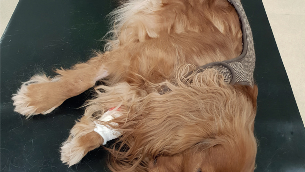
  • Post ictal — immediately after the seizure the animal may appear obtunded, disorientated or ataxic for a short period after (Figure 1).
Figure 1. This patient presented post-ictal after several cluster seizures.

A potential underlying cause of seizures, especially in dogs over 5 years of age, could be a structural finding such as a brain tumour, either primary where the tumour originates from the cells in the brain or secondary where it has spread to the brain by metastasis. The best way to confirm a brain tumour and define what type of tumour is present would be through magnetic resonance imaging (MRI) or computed tomography (CT) (Intile, n.d.).

Status epilepticus (SE)

Patients that arrive in SE can be quite worrisome to see. SE can be defined as: greater than 5 minutes of continuous epileptic seizure; or two or more discrete epileptic seizures between which there is incomplete recovery of consciousness (Berendt et al, 2015). SE is more likely to occur in dogs with structural brain disease or reactive causes of seizures (Sanders, 2015). Intravenous diazepam (Ziapam, Forum) is the most common emergency drug used in practice when attempting to stop seizure activity. This drug is highly lipid soluble which allows it to penetrate the CNS rapidly (i.e. within approximately 1 minute after intravenous administration) (Cochrane, 2007). Seizure packs are useful to attach to patients' kennels, they are pre made with the dose of diazepam on the front so it is easy to use if the patient does start to seizure (Figure 2).

Figure 2. Pre prepared seizure pack to attach to patient's kennel.

Idiopathic epilepsy

Idiopathic epilepsy (IE) is a chronic issue that can happen frequently or infrequently mainly in dogs between the ages of 1 and 5 years. If the dog is younger than 1 or older than 6, there's a 20% chance of it being idiopathic epilepsy (Gaumnitz, 2018). This is the most common type of seizure seen in practice as its exact cause is unknown, and on advanced imaging (MRI) there are no structural abnormalities found. When nursing these patients, it is important to think about things that could prevent further seizure activity, for example dimming the lights when possible, and reducing people traffic/noise in the area. Displaying warning signs on doors is an efficient way of letting all staff know there is a noise sensitive patient and to only enter if necessary (Figure 3).

Figure 3. Display warning sign to alert members of staff.

Emergency treatment

When a patient arrives at the practice seizuring, this can be distressing for both the owner and the veterinary team. Does the animal have intravenous (IV) access? This is important so antiepileptic drugs (AEDs) can be administered quickly and the patient can be administered intravenous fluid therapy (IVFT). Placing an IV catheter can be challenging due to movement of the seizuring patent, the veterinary surgeon (VS) may administer a rectal solution of diazepam (Diazepam, Desitin) to reduce the activity so IV cannulation can be facilitated. Once IV access is gained, diazepam 0.5–1 mg/kg can be administered to control seizure activity (Figure 4).

Figure 4. Emergency ready prepared catheter tray.

The duration of diazepam is short lived, so a more suitable anticonvulsant drug may then be used simultaneously. If the patient is not responding to emergency treatment and continues to seizure the animal may be set up on a constant rate infusion (CRI) of propofol (delivered using an infusion pump/syringe driver for accurate dosing) (Figure 5). Once the seizure activity has stopped, a secondary survey (full clinical examination of the patient) is done by the VS which involves checking the airway, breathing and circulation, and recording the basic but essential parameters, core body temperature, and pulse and respiration (Cortellini and De Riso, 2010). The VS should also perform a neurological examination to help them determine the origin of the seizure and obtain a baseline measurement of neurological function (Podell, 2009). This may need to be done when the effects of the propofol and diazepam have worn off.

Figure 5. Propofol constant rate infusion.

Seizuring animals will often present hyperthermic due to increased muscle activity (Lowrie, 2013). It is important to check the patient's core temperature regularly and use cooling methods when necessary, such as cool intravenous fluid therapy, laying wet towels over the patient or using fans in front of the kennel, all of which will aid in reducing temperature, at a slow and steady pace. Obtaining a blood database as soon as possible to check the parameters of glucose and calcium can assist the VS in their diagnosis and see whether the patient is hypoglycaemic due to the underlying cause or due to the intense muscular activity during the seizure. Hypoglycaemia is a common finding in seizuring animals (Cochrane, 2007a). Seizuring animals should receive 100% oxygen therapy to ensure tissue perfusion. Depending on the patient's status it can be administered via flow by, nasal prongs or cannula.

If the patient is seizuring due to a traumatic brain injury (TBI) there is a risk of oedema therefore potentially causing increased intracranial pressure (ICP). Once the seizures are controlled, the VS may opt to give the patient a hyperosmolar fluid agent, such as mannitol or hypertonic saline.

These fluids work by increasing the osmotic gradient across the blood brain barrier (BBB), and draw fluid out from the cells into the vasculature, which reduces the ICP in the brain. ICP can be recognised by the Cushing's Reflex. This reflex is classically seen as an increase in blood pressure, bradycardia and an irregular respiratory rhythm. If a Cushing's reflex was to occur there is high chance of the brain herniating resulting in death. As a result, a Cushing's reflex suggests a need for immediate care as its presence is a good indicator of high ICP (Ayling, 2002).

Clinical history

Initial consent from the owner should be obtained as soon as possible so the team can work to stabilise the patient while a colleague can then collect a full clinical history (Bloor, 2013). When presented with a patient who is/was seizuring the top three objectives are:

  • To stop the seizure
  • To find out the possible cause for the seizure
  • To confirm that the episode was a seizure.

VSs rely on the owner's interpretation of any potential ‘seizure activity’ to initially diagnose epilepsy; because there are many ways dogs may act during a seizure, some owners are not actually aware their dog had or is having a form of seizure. Therefore, home video recordings are very useful for the VS to determine whether a seizure has occurred or not (Vite and Long, 2007), particularly if the owner's description is insufficient.

First of all, obtain the patient's signalment, i.e. species, breed, weight, age, gender. Find out if the patient is on any current medication or has any other medical conditions. In addition, also determine if there is any potential risk the patient may have ingested something toxic (e.g. ethylene glycol from antifreeze may cause seizures up to 12 hours after ingestion (Pet Poison Helpline, 2018). Finding out if the patient has shown any signs of seizures in the past, or if the animal has had any behavioral changes (Bloor, 2013). The information gathered should be reported to the VS in charge to guide the management of the patient and aid any further work up.

Diagnosis

Blood work such as a haematology and biochemistry is important to collect on presentation to help rule out a metabolic origin. If there is suspicion of an intracranial cause, then MRI can be performed under general anaesthesia, but is important to stabilise the patient prior to imaging so they are a safe anaesthetic candidate. While the animal is under general anaesthesia a cerebral spinal tap can be undertaken, this is an essential component in the investigation of neurological diseases; cerebrospinal fluid (CSF) is taken at the cerebellomedullary cistern using a spinal needle. Correct patient positioning is key to achieving a successful tap. Some CSF analysis results support specific diagnoses (Vite and Long, 2007). Diseases in the CNS often reflect in the CSF when the BBB is compromised — the liquid should be clear/transparent with a low protein, cell numbers often will increase in acute inflammation and some other conditions (Braund et al, n.d.).

The decision to start any antiepileptic drugs (AEDs) depends on the individual patient. AEDs should generally only be started if a single seizure occurs more than once every 4–6 weeks, cluster seizure activity or status epilepticus regardless of frequency (Cochrane, 2007b).

Long-term treatment

The animal may remain on AEDs for the foreseeable future. Educating the owner on the drugs, you are giving, and possible side effects are crucial to ensure the medication is effective. There are several drugs, which may be considered:

  • Phenobarbital (Epiphen, Vetoquinol; EpiRepress, Virbac) requires regular blood testing to measure the amount of the drug in the animal's blood to ensure the administered dose is controlling the seizures. Be aware long-term use of phenobarbital has been reported to cause liver damage in dogs (Muller et al, 2010).
  • Potassium bromide (KBr) is the oldest of anticonvulsant drug used in dogs, it works by replacing chloride in all body fluids and stabilises neuronal cell membranes by impeding chloride transport and activating the effect of gamma-aminobutyric acid (GABA) (Dowling, 1994). A disadvantage to using KBr is that it may take up to 16 weeks to reach a steady serum level (Bagley and Chen 2013).
  • Imepitoin (Pexion, Boehringer Ingelheim) was licensed in 2013 and is used to reduce the frequency of seizures, it also reduces the anxiety and fear assosicated with noise phobia. Studies show dogs treated with imepitoin developed cluster seizures more frequently then those treated with phenobarbitone (Stabile et al, 2019). It has been recommended to use imepitoin in dogs experiencing recurrent single generalised epileptic seizures (Bhatti et al, 2015).
  • Levetiracetam (Keppra, UCB) is an AED licensed for use in humans. It appears to be relatively safe in dogs, although studies are still ongoing to investigate any adverse effects.

While treatment may be with a single drug only, it is suggested that up to 50% of dogs will require multiple AED therapy (Cochrane, 2007a). Ensuring the patient is receiving the medication at the same time each day is foremost for maximum efficiency — some oral medications require twice daily administering (BID), or three times daily (TID). The owner should also be made aware that this is long-term treatment for their pet, which can become very costly, especially if the pet is not insured (Figure 6).

Figure 6. Some of the commonly prescribed antiepileptic drugs (AEDs) used to treat seizures in canine patients.

The Royal Veterinary College (RVC) has developed the first app for pet owners to monitor and record seizure activity from their mobile phone or tablets. The RVC Pet Epilepsy Tracker helps owners increase their knowledge should their pet be diagnosed with epilepsy. It also includes reminders when medications are due to be given if the animal is on multiple daily medications, as many dogs with the disease are on multiple anticonvulsant drugs at one time (Woodbine, 2015).

Conclusion

Dealing with emergency seizuring patients can be a stressful time for all involved. There is not one set protocol to follow when dealing with a seizuring patient, treatment is based on each individual and how they present. The veterinary nurse must be aware of their role in the management of seizuring patients, understanding the patient's requirements and nursing through the subsequent monitoring period. It is important to remember epileptic seizures cannot always be cured; AEDs simply control the disease, and it is important to keep in mind the animal's quality of life.

KEY POINTS

  • Seizures can be split into two classifications intracranial and extracranial.
  • Three stages of generalised seizures are pre-ictal, ictal and post ictal.
  • Clincal signs can vary from full tonic clonic to focal seizures where twitching, hypersalivating or head shaking can be observed.
  • Seizures especially status epilepticus can be potentially life threatening.