The veterinary nurse's role in the management of acute oropharyngeal injury in dogs
02 June 2022
13 mins read
Volume 13 · Issue 5
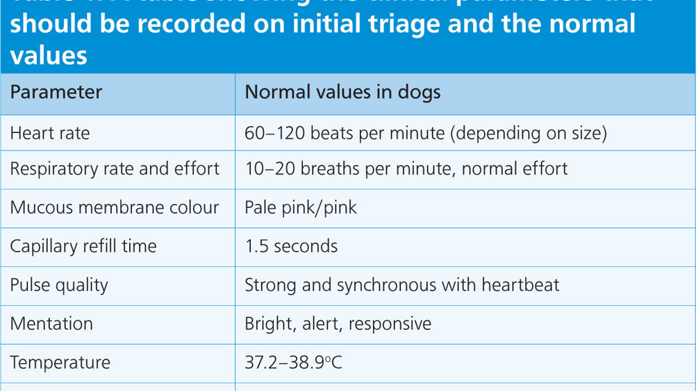
Table 1. A table showing the clinical parameters that should be recorded on initial triage and the normal values

Abstract

Oropharyngeal injuries are commonly seen in practice. Severity can range from minor the life threatening. It is important that veterinary nurses can confidently perform an initial triage, recognise life-threatening problems, and provide initial stabilisation to these patients. Nurses play a fundamental role in the management of these cases throughout their stay in hospital. This article aims to provide practical advice and guidance on cases of oropharyngeal injury, and the importance of client education to help prevent similar cases in the future.

Acute oropharyngeal puncture wounds are relatively uncommon in general practice but are potentially life-threatening events if not recognised promptly (Griffiths et al, 2000). In cats and dogs, they can be caused by a variety of foreign bodies such as wood, metal, bones, sewing needles and fishhooks (White and Lane, 1988; Griffiths et al, 2000). In dogs, the most common presentation is associated with carrying, chewing, or retrieving wooden sticks (Hallstrom, 1970; White and Lane, 1988; Doran et al, 2008; Anderson, 2017).

Two presentations of oropharyngeal injury are typically seen clinically; the acute and chronic stages (Griffiths, et al, 2000; Doran et al, 2008). Acute injury is defined as cases that present within 7 days of the traumatic event, whereas chronic cases are those presenting after this time (Baker, 1972; White and Lane, 1988). This article aims to provide practical advice and guidance to veterinary nurses managing cases with acute oropharyngeal disease.

Triage

A successful outcome of an emergency patient is more likely where quick and appropriate action is taken. This is dictated by the initial observation and clinical examination. Triage is a way of rapidly evaluating patients and providing treatment to those that are the most urgent (Aldridge and O'Dwyer, 2013). In an individual case, it is allocating treatment to the most serious problem first. In cases of oropharyngeal stick injuries, a wide range of problems may present. Presenting signs in acute cases are often a combination of cervical pain, pain on flexion of the neck, gagging, hypersalivation, blood-tinged saliva, dysphagia, and depression (Doran et al, 2008; Robinson et al, 2014; Anderson, 2017).

A quick capsular history should be obtained from the owner on arrival at the clinic. This includes asking the signalment (age, sex, neutered, breed), what the presenting problem is, the duration of the presenting complaint and any current medications (Boag and Marshall, 2020). Owners may recognise a specific event leading to the injury, such as their dog running for or retrieving a stick (Robinson et al, 2014). Alternatively, owners may be out of sight from the event and the presenting signs might raise suspicion of an oropharyngeal stick injury.

Examination of the major body systems should be carried out, which includes the respiratory, cardiovascular, and central nervous system (CNS). These three systems should always be examined first, regardless of other injuries (Boag and Marshall, 2020). Initial examination should concentrate on a small number of physical parameters to provide the most important information (Aldridge and O'Dwyer, 2013). Parameters that should be recorded and the normal values in dogs are seen in Table 1. A blood sample should be obtained for a minimum database (packed cell volume/total solids, electrolytes, blood glucose and lactate). This will complement the initial examination and act as a baseline to monitor trends while the patient is hospitalised.


Table 1. A table showing the clinical parameters that should be recorded on initial triage and the normal values
Parameter Normal values in dogs
Heart rate 60–120 beats per minute (depending on size)
Respiratory rate and effort 10–20 breaths per minute, normal effort
Mucous membrane colour Pale pink/pink
Capillary refill time 1.5 seconds
Pulse quality Strong and synchronous with heartbeat
Mentation Bright, alert, responsive
Temperature 37.2–38.9oC
Gait Ambulatory

Stabilisation

It is important that any abnormal changes on initial examination are addressed. Any patient with an oropharyngeal injury will require a general anaesthetic. Reasons for this are to assess the extent of any injuries, to protect the airway, provide ventilatory support and to provide a motionless surgical field if required. By stabilising patients prior to the induction of anaesthesia, there is less chance of the patient decompensating and deteriorating (Aldridge and O'Dwyer, 2013). The temptation to rush unstable patients to anaesthesia must be avoided.

Respiration

Respiratory rate, pattern and effort should be assessed initially before handling the patient. Auscultation of the patient's chest should be performed, which will provide information on cardiac and respiratory function (Farry and Norkus, 2018). Cardiac arrythmias, murmurs or dull/absent heart and lung sounds, all well as increased pulmonary noise such as wheezes/crackles could indicate abnormalities (Aldridge and O'Dwyer, 2013). If there is any concern regarding cardiac or respiratory function, then further evaluations and testing should be performed. Oxygen therapy may be indicated where pharyngeal swelling or mediastinal emphysema causes dyspnoea (Reineke et al, 2014). In cases with severe pharyngeal swelling or laryngeal trauma, a temporary tracheostomy tube may be required (White and Lane, 1988).

Cardiovascular system

Assessment of the cardiovascular system (CVS) provides the veterinary nurse with information about a patient's ability to perfuse tissues adequately. Evaluating mucous membrane colour, capillary refill time, pulse rate, rhythm and quality alongside thoracic auscultation and arterial blood pressure measurements, provides a baseline of the patient's cardiovascular function (Aldridge and O'Dwyer, 2013). Prior to the induction of anaesthesia, an intravenous catheter should always be placed, and aggressive intravenous fluid therapy may be indicated to correct hypovolaemia (Doran, et al, 2008). Anaesthetic drugs are likely to result in significant depression of the CVS in critically ill patients, which will result in a further reduction in the system's ability to perfuse tissues and meet oxygen demands (Aldridge and O'Dwyer, 2013).

Neurological system

Patients with oropharyngeal stick injuries may present with many variations in neurological status, ranging from lethargy, obtundation, stupor or coma (Farry and Norkus, 2018). Nurses should inform the veterinary surgeon of any deterioration in the ambulation and neurological status of a patient as further work-up and treatment may be required (Aldridge and O'Dwyer, 2013).

Investigation

Anaesthesia

For patients with oropharyngeal wounds, general anaesthesia will be required prior to further investigations. These patients require careful assessment and planning of anaesthetics to minimise complications and ensure a safe anaesthetic (Murrell and Ford-Fennah, 2020). The goal of anaesthesia in any critical patient is to maintain tissue and organ perfusion and ensure sufficient oxygen delivery and carbon dioxide removal.

The goal of premedication in these patients will be to provide analgesia, promote sedation and amnesia, and to reduce the dose of induction agent required (Leonard, 2017). Full mu-opioids are frequently used in critical patients to provide excellent analgesia, while having minimal effect on the cardiopulmonary system and CNS (Murrell and Ford-Fennah, 2020). Opioids may be used alone or can be combined with tranquillisers, to produce neuroleptanalgesia or with sedatives to produce increased sedation (Aldridge and O'Dwyer, 2013). Classes of drugs such as alpha-2-agonists and phenothiazines should be avoided in these patients because of their cardiopulmonary effects. Benzodiazepines cause anxiolysis, sedation and muscle relaxation with minimal cardiopulmonary effects, however, their analgesic effects are minimal, so they are often combined with opioids for sedation (Murrell and Ford-Fennah, 2020). Alternatively, they can be used as a co-induction to reduce the amount of induction agent required to produce a loss of consciousness (Aldridge and O'Dwyer, 2013). Most of the commonly used induction agents (propofol and alfaxalone) cause hypotension even in healthy patients, and this can be profound in critical patients (Aldridge and O'Dwyer, 2013). Patients suffering from trauma should be expected to have a full stomach, so rapid intubation to protect the airway is important. Pre-oxygenation is recommended prior to induction of anaesthesia, to increase the partial pressure of oxygen within arterial blood (PaO2) (Leonard, 2017). This will reduce the chance of hypoxia and desaturation during induction (Pascoe, 2011).

Electrocardiography (ECG), capnography and arterial blood pressure should be monitored during induction, so that acute changes associated with anaesthetic agent administration can be identified rapidly and appropriate action taken. A critical patient will often require a lower vaporiser setting than a healthy patient (Fletcher, 2014). Therefore, after induction, the patient's depth of anaesthesia should be assessed by looking at their eye position, jaw tone, and assessing whether a palpebral reflex is present. The cardiovascular function can be assessed by listening to the heart, palpating peripheral pulses, using pulse oximetry and capnography. Table 2 describes patient parameters that should be monitored during general anaesthesia, normal values, and ways of monitoring them. It is important to record every parameter every 5 minutes, as well as recording all drugs and dosages administered (Leonard, 2017).


Table 2. Patient parameters that should be regularly monitored during general anaesthesia, normal values, and different ways to monitor them
Parameter Normal values in dogs Monitoring
Heart rate 80–120 bpm Auscultation (transthoracic, oesophageal), pulse palpation (rate, rhythm, quality), ECG
Blood pressure 120/80 (100) mmHg Minimum acceptable value 80/40 (60) mmHg Pulse palpation (rate, rhythm, quality), blood pressure (direct or indirect (Doppler, oscillometric))
Ventilation ETCO2 35–45mmHg RR 10–20 breaths per minute Movement of chest/circuit bag, capnography (ETCO2), arterial blood gas
Oxygenation SpO2 >96% Mucous membrane colour, pulse oximetry, arterial blood gas monitoring
Temperature 37.2–38.9oC Rectal or oesophageal temperature probe

bpm = beats per minute; ECG = electrocardiogram; ETCO2 = end tidal CO2; RR = respiratory rate; SPO2 = saturation of peripheral oxygen

Assessment of the oropharynx and imaging

Following induction of anaesthesia and intubation, a thorough inspection of the oral cavity and pharynx must be performed (White and Lane, 1988; Fossum, 2018). Equipment such as long bladed laryngoscopes, tongue depressors and a good light source will be required for an effective oral examination (Doran et al, 2008) (Figure 1). Orthogonal radiographs of the pharynx, cervical region and thorax should be obtained (Anderson, 2017; Fossum, 2018). If endoscopy is available, the oesophageal lumen can be evaluated (White and Lane, 1988; Huck and Kyles, 2017).

Figure 1. Laryngoscopes, blunt probes and surgical light sources are used to conduct a thorough examination of oropharyngeal wounds.

Advanced imaging such as computed tomography (CT) and magnetic resonance imaging (MRI) have not been evaluated for their use in acute oropharyngeal injury. CT and MRI have been shown to be useful in chronic cases (Dobromylskyj et al, 2008; Nicholson et al, 2008; Anderson, 2017). CT can be useful for identifying wooden foreign bodies because of its removal of superimposition and cross-sectional nature when compared with radiography (Lamb, et al, 2017).

Treatment

In dogs with simple, acute, rostral oropharyngeal penetrations with no radiographic abnormalities, an oral approach is used to manage the wounds (Doran et al, 2008). Wounds should be probed and explored to assess the depth of penetration and to remove any residual foreign material present (Griffiths et al, 2000; Doran et al, 2008; Fossum, 2018). The pharynx should be packed before copious lavage with warm lactated ringers’ solution to minimise any residual contamination and dislodge any remaining foreign material (White and Lane, 1988; Anderson, 2017).

Surgical treatment

Where investigations of these cases lead to a suspicion of caudal pharyngeal or oesophageal damage, surgical exploration of the cervical region should be performed (Figure 2). A ventral cervical midline approach is taken. If a contaminated wound has been identified, antibiotic prophylaxis is indicated prior to surgery (Anderson, 2017; Fossum, et al, 2018).

Figure 2. Intraoperative view of a ventral cervical exploration demonstrating a single puncture wound of the dorsal oesophageal wall. Orientation: cranial (left of image).

The patient should be positioned in dorsal recumbency. An extensive clip of the neck is made from the chin (cranial) to the xiphoid process (caudal) and extended laterally both sides. Loose hair should be vacuumed or placed in a bin. Skin preparation should use a 50:50 dilution of chlorhexidine gluconate with warm water and sterile swabs. Gauze swabs are more suitable as cotton wool tags can adhere to the skin stubble (Whiting et al, 2006). Scrubbing should commence at the intended incision line moving in an outward direction. Oral preparation can also be performed using a chlorhexidine gluconate dilution of 1:80.

Appropriate instrumentation will facilitate successful cervical exploration. A soft tissue pack should include a scalpel handle, towel clamps, a range of haemostatic forceps, needle holders, Mayo and Metzenbaum scissors (straight and curved) and thumb forceps. A range of retractors should be provided (see Figures 3 and 4). Additionally, active closed suction drains, such as Jackson-Pratt drains, should be prepared. Placement of a surgical drain is indicated to reduce dead space and any residual contamination (Figure 5). Oropharyngeal bypass can be performed if deemed necessary by the surgeon. A variety of feeding tubes should be prepared prior to surgery, e.g. naso-gastric, oesophageal and gastrotomy tubes. The choice of feeding tube will depend on the patient, however gastrotomy tubes are recommended for postoperative nutritional support and to provide medications in patients with oropharyngeal stick injuries (White and Lane, 1988; Doran et al, 2008; Fossum et al, 2018).

Figure 3. A range of handheld retractors. 1: Weislander 2: Malleable 3: Hohman 4: Langenbeck 5: Gelpi.
Figure 4. Intraoperative view of a ventral cervical exploration. A ring retractor has been used to retract the paired sternohyoid muscles from the midline. Orientation: cranial (left of image).
Figure 5. Placement of an active closed suction drain following cervical exploration.

Postoperative management

Close monitoring will need to continue in the postoperative period. The parameters used to record the initial triage should be used to monitor the critical patient after investigations and/or surgery (Table 1). Consistent, regular, and reliable monitoring by the veterinary nurse is fundamental in the monitoring of the critical patient (Farry and Norkus, 2018). Hypothermia is a common yet preventable side effect of anaesthesia and surgery. Prolonged abnormal body temperatures interfere with a patient's homeostatic mechanisms and delay return to normal health. Where active warming is required in hypothermic patients, ensure care is taken not to cause overheating or local burns (Aldridge and O'Dwyer, 2013). Ensure that adequate analgesia is provided in the postoperative period. Regular pain scoring using a reliable scoring method will indicate how painful a patient is and whether an increase or reduction in analgesia is required. There are many reliable pain scoring methods available for use in veterinary practice. The Glasgow Composite Measure Pain Scale (GCMPS) and its short form (GCMPS-SF) are validated multi-dimensional pain scales and are frequently used in veterinary practice (Matthews et al, 2014). Consideration must be made to continually re-assess and replace fluid as a result of preexisting fluid deficits or vascular volume expansion associated with anaesthetic agents used (Aldridge and O'Dwyer, 2013).

Management of drains

Placement of a surgical drain is indicated to reduce dead space and any residual contamination. Active closed suction drains are more suitable than passive drains in the cervical region, as they do not require a dependent exit point or repeat bandaging (Anderson, 2017). Active closed suction drains require active removal of fluid using suction (Whiting et al, 2006). For this to be possible, the drain must be airtight. A collection chamber is used to achieve this. To activate the drain, the chamber is opened and squeezed to remove air, then closed. The chamber will then return to its original shape and suction is generated to draw out fluid and exudate from the wound.

Drains should be monitored closely to identify complications early and ensure they are functioning correctly. Wounds with surgical drains should be monitored for signs of surgical site infections (SSI). Signs include exudate, erythema, oedema, pain, pyrexia, lethargy and depression (Yon, 2020). The risk of ascending infections with active closed suction drains is present, however it is considered much lower than open passive drains (Reifell et al, 2013). When handling drains, ensure an aseptic technique is used. Drains should be dressed to provide a protective barrier between the wound and external environment. Strikethrough of exudate may occur which can facilitate bacteria passing through the dressing to contaminate the wound underneath. Dressings must be changed before this is observed (Gray, 2018). Changing dressings too frequently may increase the risk of contamination and ascending infections if not handled aseptically. Veterinary nurses must therefore monitor dressings and change them when deemed necessary (Yon, 2020).

Patient interference can result in premature removal of drains or SSIs; therefore, it is vital to ensure adequate techniques are used to prevent self-removal or interference (Yon, 2020). In cases of oropharyngeal stick injuries with cervical drains present, body suits or netting over bandaging may be useful. Elasticated netting can also be useful to position the collection chamber of a drain to avoid it creating excess tension on the wound. Placement of drains may lead to local inflammation as a result of the body recognising it as foreign material (Watson and McFadden, 2019). This may lead to pain and discomfort in addition to that caused by the initial oropharyngeal injury or surgical incision. It is therefore essential that regular pain scoring is performed, and that adequate analgesia is provided to maintain comfort and decrease the chance of self-trauma or removal of the drain.

Surgical drains should be monitored, and outputs should be recorded. Removal is performed when fluid recovery falls to an acceptable level (less than 0.5 ml/kg in (Doran et al, 2008)). The longer the drain is left, the greater the risk of bacterial colonisation at the drain site (McFadden and Oberhaus, 2019). However, if a drain is removed too early, the risk of seroma formation is increased (Shaver, et al, 2014).

Management of feeding tubes

Nurses managing feeding tubes should be happy with the type of tube, how to administer foodstuffs and the type of foodstuff that is suitable to be administered. It is likely that a gastrotomy tube will be placed to bypass the oesophagus if cervical exploration has been performed (Figure 6). Prior to feeding, integrity of the tube needs to be assessed and all connectors should be checked for damage. The stoma site and dressings should be checked, and hospital records checked to see whether a clean or a dressing change is due. Dressing changes should occur at least once a day (Lumbis, 2017). Oral nutrition can be offered after 7 days to allow adequate time for the oesophagus to heal. Tube feeding can continue if the patient is reluctant to eat. Tubes should be suctioned with a syringe to check for large gastric residual volumes. Next, the tube should be flushed with water prior to feeding to check it is patent and in the correct position. Feeding can then commence.

Figure 6. Gastrostomy tube surgically placed via a flank approach. Orientation: cranial (left of image).

Gastrostomy tubes should be left in situ for a minimum of 10 days to allow for the formation of adequate adhesions between the stomach and body wall (Davidson, 2017). Gastrostomy tubes should be left in until enteral feeding is no longer required (Davidson, 2017). Doran et al (2008) fed cases on day 10 postoperatively, and if there was no evidence of dysphagia or regurgitation, the tubes were removed on day 11.

Client education

Education of clients by veterinary professionals plays an important role in improving animal welfare. Cases of oropharyngeal injury by sticks are challenging to manage, yet preventable, therefore it is important that owners are well educated about the risks. In one study, only 12% of cases were presented to a veterinary practice within 7 days of injury despite 48% of all the cases having a known history of oropharyngeal penetrating trauma (Griffiths et al, 2000). There are many ways to educate clients on important topics such as stick injuries. First, eye catching displays or posters with simple and to the point information are an easy and cheap way to catch a client's attention. Similar posters and pictures can be shared on social media platforms to attract the attention of clients that are not at the practice, or for people who are not clients but follow the practice online. Nurse clinics and puppy classes are an excellent way to ensure clients are educated properly on preventative health care. They allow clients to be taught by veterinary professionals on relevant topics. They also allow clients to ask questions in person and obtain reliable answers.

KEY POINTS

  • Acute oropharyngeal injury is uncommon in first-opinion practice but potentially life threatening.
  • A successful outcome of an emergency patient is more likely where quick and appropriate action is taken. Initial triage is important to recognise life threatening problems and to provide stabilisation.
  • General anaesthesia is often required to assess the oropharynx. These patients require careful assessment and planning of anaesthetics to minimise complications and ensure a safe anaesthetic.
  • Some cases require surgical exploration of the cervical region. Appropriate instrumentation will facilitate this.
  • Close monitoring will need to continue in the postoperative period where cervical exploration has been performed and nurses should be confident managing drains and feeding tubes.

Conclusions

Cases of acute oropharyngeal injury are challenging yet preventable and potentially life threatening. These cases may present with a variety of clinical abnormalities. Initial triage and stabilisation of abnormal changes is important prior to general anaesthesia to prevent decompensation and deterioration. The literature shows a benefit from an aggressive and thorough approach to such injuries on initial presentation. Multiple methods can be used to manage these injuries including endoscopic debridement and cervical exploratory surgery. The choice of drugs for anaesthesia should be chosen on an individual basis depending on how the patient presents itself. Close monitoring in the postoperative period is important and nurses should be confident managing drains and feeding tubes. It is important that measures are taken to educate clients on the risks of sticks to prevent future cases.