A patient care report of a Doberman in heart failure
01 November 2011
9 mins read
Volume 2 · Issue 9
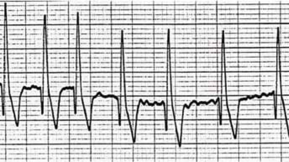
Figure 1. Electrocardiogram (ECG) showing atrial fibrillation in a Doberman. Heart rate is between 220–250 beats/min, note the lack of P waves and irregular rhythm. ECG courtesy of Simon Dennis.

Abstract

This article describes the nursing care provided to a Doberman in acute life-threatening heart failure due to dilated cardiomyopathy (DCM). DCM is a common problem seen in medium-large breed dogs. It can sometimes lead to congestive heart failure (CHF) and cause arrhythmias, further compromising cardiac function. Nursing care, monitoring and therapy are vital for the patient both in the short term, but also long term, to optimize quality of life.

Signalment:

Species: Canine

Breed: Doberman

Age: 7 years

Sex: Male (neutered)

Weight: 43.3 kg

Reason for admission

Henry was presented with acute onset respiratory distress. He had a 2 month history of exercise intolerance and breathlessness, but had developed a retching cough 4 days prior to presentation, and had experienced one syncopal episode.

Patient assessment

On presentation, the patient had a frail, quiet but alert demeanour. He had audible crackles over his lung fields, a respiration rate (RR) of 40 breaths/minute with increased respiratory effort (RE), and a cough. He had a grade 2/6 left-sided, apical, systolic heart murmur and weak peripheral pulses. His heart rate was 140 beats/minute, with no pulse deficits. Temperature was 38.2°C.

Initial diagnostics and results

An intravenous (IV) catheter was placed in a cephalic vein, and blood was taken for haematology and biochemistry from the catheter. A mild hyperkalaemia (4.78 mmol/l), and mild hyperglycaemia (6.78 mmol/l) were the only abnormalities found. On the basis of his history and physical examination, congestive heart failure (CHF) was suspected. The veterinary surgeon instructed IV furosemide (2 mg/kg) to be given, and the patient was settled into a kennel with supplemental oxygen provided by nasal prongs. A continuous electrocardiogram (ECG) recorded a sinus rhythm with an average heart rate of 150 beats/minute. He was allowed to adjust to his new surroundings before further diagnostic tests were performed.

About an hour later, echocardiography showed a rounded, poorly contracting left ventricle with atrial dilation. Thoracic radiographs were consistent with cardiogenic pulmonary oedema. Systolic blood pressure was 100 mmHg. The assessment confirmed that Henry had left-sided congestive heart failure (L-CHF), which was secondary to dilated cardiomyopathy (DCM). His blood pressure was borderline low, possibly refecting low output heart failure.

Medical management

The veterinary surgeon started a treatment plan for Henry. This included:

  • Furosemide (2 mg/kg), given twice at 1–2 hourly intervals (after initial dose on administration). The dose was then decreased to (2 mg/kg) IV every 3 hours. The dose and timing was based on monitoring of RR and RE
  • A dobutamine infusion started at a dose rate of 5 μg/kg/minute. This was administered for 12 hours.
  • Pimobendan per os twice daily (0.2 mg/kg)
  • Glyceryl trinitrate ointment 1 inch on alternate pinnae four times daily.


Initial nursing intervention

Monitoring of the patient in CHF is essential, however, it must never compromise the patient. Stress should be avoided in the CHF patient because many patients have respiratory impairment due to pulmonary oedema, pleural effusion, or severe ascites. When the patient presents in CHF, the cardiovascular system is already working to capacity, trying to maintain adequate blood pressure in response to the reduced cardiac output from heart disease. Any extra stress or exertion could precipitate a life threatening arrhythmia. For an anxious patient in respiratory distress, monitoring RR and RE from a distance may be all that is possible. Obtaining IV access is highly desirable because it is the most efficient route for drug therapy to take maximal effect. However, some patients may not be stable enough initially to allow placement of an IV catheter, particularly if they are distressed. If the patient tolerates it, regular heart rate and rhythm checks are also important. This is preferably done with a continuous ECG, which should minimize handling and subsequently stress, and allows detection of arrhythmias. Accurate and reliable blood pressure measurement is also useful as this provides information to assess whether the patient is becoming hypotensive. This should be performed with minimal handling and stress to the patient.

The immediate nursing plan for Henry was:

  • Observation and recording of RR and RE hourly to monitor effects of drug therapy
  • Monitor heart rate and rhythm hourly
  • Provide and maintain oxygen therapy
  • Keep clean and comfortable
  • Ensure fresh water is available at all times
  • IV catheter management. A small volume of dilute heparinized saline was flushed every 4 hours to maintain catheter patency. The catheter was undressed and inspected daily for signs of inflammation and swelling. It was removed as soon as was possible
  • Give medication accurately and on time
  • Monitor demeanour hourly
  • Monitor systolic blood pressure twice daily
  • Ensure that all staff are aware to keep the patient calm and exercise restricted.


Ongoing patient assessment

Henry responded very well to medical therapy. His RR and RE were within normal limits the next day. He was taken on restricted walks around the intensive care unit the next day. He slowly regained his appetite and started eating when hand fed. Medications were changed from IV to oral, and he was allowed to go home on day three of hospitalization. He was sent home with the following medications:

  • Furosemide 100 mg every 8 hours per os (2 mg/kg)
  • Pimobendan 10 mg every 12 hours per os (0.2 mg/kg)
  • Spironolactone 100 mg every 24 hours per os (2 mg/kg)
  • Benazepril 20 mg every 24 hours per os (0.4 mg/kg)


Henry was re-examined on two further occasions. At the first appointment (7 days later), his heart rate was 160 beats/minute and weak femoral pulses were noted on physical examination. The owner reported that Henry had been bright at home, with a RR between 20–25 breaths/minute. Blood tests showed a mild increase in urea, while creatinine and electrolytes were all within normal parameters. Systolic blood pressure was 115 mmHg and thoracic radiographs showed a continued improvement in pulmonary congestion. On his second re-examination (21 days later), physical examination findings showed that he still had weak femoral pulses with a pulse rate of 150 beats/minute. The owner was still pleased with his progress (RR still between 20–25 breaths/minute) and had reduced the furosemide from 100 mg to 80 mg every 8 hours. Repeat thoracic radiographs showed that the pulmonary congestion had cleared and heart size had decreased. Blood tests for urea, creatinine and electrolytes were within normal limits except for a mild hyponatremia. The furosemide dose was decreased again to 80 mg every 12 hours. His next examination has been scheduled for 2 months' time.

Ongoing nursing intervention

When Henry came back for re-examination after being diagnosed with CHF, it was still important to minimize stress. A quiet room was used for consultation and physical examination. The owners were questioned as to what the RR was at home, and how he coped with exercise. They were also asked about appetite, demeanour, urination and drinking habits. Henry was weighed on every visit to the hospital. Monitoring of body weight is important because it can be indicative of anorexia, but also fluid retention. If ascites is present, measuring abdominal girth should be considered.

Case discussion

DCM is the second most common acquired cardiac disease in the dog (O'Grady et al, 2008). DCM is over-represented in certain breeds, including Dobermans, Newfoundlands, Irish Wolfhounds and Great Danes, and is thought to be a hereditary disease in many breeds (Ware, 2007). Cardiomyopathy is a term used to describe an inadequately functioning heart muscle, which means that the ventricle fails to expel all the blood in systole, leading to progressive dilatation of the ventricle. This incompetency will eventually lead to CHF. The dilatation of the ventricle also distorts the mitral valve apparatus, which in turn, increases the potential for heart failure due to increased pulmonary venous pressure and/or atrial arrhythmias.

Some dogs remain in sinus rhythm, but often tachyarrhythmias such as atrial fibrillation can occur. Ventricular arrhythmias may also be seen, and the presence of a complex ventricular arrhythmia, such as ventricular tachycardia, may be associated with an increased risk of sudden death (see Figures 1 and 2). The two main reasons for death in Dobermans with DCM are sudden death and euthanasia because of refractory CHF. It has been shown that Dobermans commonly present with ventricular arrhythmias, and this has been strongly linked to the higher sudden death rate in this breed (Calvert et al, 2000). Prognosis for Dobermans presenting with CHF is guarded to poor, however a favourable response to initial treatment is a good prognostic indicator (Sisson et al, 1995).

Figure 1. Electrocardiogram (ECG) showing atrial fibrillation in a Doberman. Heart rate is between 220–250 beats/min, note the lack of P waves and irregular rhythm. ECG courtesy of Simon Dennis.
Figure 2. Electrocardiogram (ECG) showing intermittent ventricular tachycardia in a Doberman. Note the sinus complexes (numbers 1, 17, 20). The other complexes are ventricular in origin, and regular in appearance. The ventricular rate is 300 beats/min. This can be a life-threatening arrhythmia because the rate can be so fast that the heart does not have time to repolarize (and subsequently refill) before it beats again. ECG courtesy of Simon Dennis.

Dogs with DCM may present with vague clinical signs, i.e. cough, exercise intolerance, weight loss, or they can present with acute, life-threatening CHF as happened in this case. Clinical signs of acute CHF include respiratory distress, cough (sometimes producing pink froth), and collapse. In patients with DCM, cardiac output is compromised, arterial blood pressure falls, which leads to the activation of compensatory mechanisms, in an attempt to return blood pressure to a more normal level. These compensatory mechanisms, namely the sympathetic nervous system and the renin-angiotensin-aldosterone axis, increase fluid retention by the kidneys, vasoconstriction, and stimulation of the heart to increase heart rate and stroke volume (Oyama, 2010). Although ultimately beneficial to restore adequate arterial pressures, the ‘trade-of’ is an increase in venous pressures, which if severe enough can cause fluid to leak (effuse) into the surrounding interstitial tissues (pulmonary oedema) or cavities (ascites, pleural effusion), resulting in CHF. If arterial pressure cannot be normalized despite maximal activation of compensatory mechanisms, then low output heart failure is present. Affected patients usually have concurrent CHF, unless the fall in output is so acute that compensatory mechanisms have insufficient time to be activated (e.g. some arrhythmias, myocardial infarction) (Dennis, 2010; Oyama, 2010). Although life saving in the short term, long-term stimulation of compensatory mechanisms causes progressive deterioration of cardiac function due to fatigue of the heart muscle. Anorexia and cardiac cachexia are acknowledged complications of heart failure. One study showed that anorexia was a contributing factor in an owner's decision to euthanize their pet with CHF in 68% of cases studied (Mallory et al, 1999).

Nursing care

Correct and timely nursing intervention is extremely beneficial for the patient in CHF. Some of the key points that the role of the nurse should include are included in Table 1.


Table 1. The key role of the veterinary nurse
General management
Liaise and educate the owner of the clinical signs to monitor, such as tachypnoea, breathlessness (not panting), increased resting RE, exercise intolerance and anorexia
Ensure appetite remains good, and discuss alternatives with the owner if the patient is not eating. Dietary supplementation of Omega-3 fatty acids has been shown to be beneficial in patients with CHF (Freeman, 2010)
Acute management
Provide supplemental oxygen therapy
Keep stress to a minimum for the patient
Monitor respiratory rate (RR) and respiratory effort (RE)
Keep patient clean and dry, especially if exercise is restricted
Maintain intravenous catheter access
Calculate, administer and monitor drug therapy
Assist with diagnostic procedures
Prepare kits for thoracocentesis/abdominocentesis
Chronic management
Monitor body condition
Monitor changes in demeanour
Monitor blood pressure (record how the procedure was done, so that circumstances can be repeated on each occasion)
Apply 24 hour electrocardiogram (ECG) (Holter) monitors if required, to monitor heart rate and rhythm at home
Assist with diagnostic procedures
If ascites is present, monitor changes in body weight and measure abdominal girth

It is also important that nurses understand the drugs that are used in the management of heart failure, because it will help their understanding of heart disease and potential side effects. Henry was given immediate treatment on admission to the hospital. Furosemide was given because it is a fast and potent diuretic, with maximum effect 20 minutes after IV administration. He was also given dobutamine, which is a synthetic catecholamine that has a positive inotropic effect. Positive inotropes increase the strength of the ventricular contraction, thus improving cardiac output. Dobutamine is usually reserved for patients with acute life-threatening heart failure, and must be given as a slow infusion on a short-term basis (for no longer than 48–72 hours) as it loses efficacy over time. A continuous ECG must be used because the adverse effects of dobutamine include ventricular arrhythmias and/or tachycardia. Dobutamine can also cause hypokalaemia as a result of potassium uptake into cells. For additional and longer-term inotropic support, Henry was given pimobendan, which has both positive inotropic and vasodilatory effects. One study found that pimobendan increases longevity in Dobermans with CHF (O'Grady et al, 2008). Glyceryl trinitrate was applied to the pinnae because it is a systemic venodilator that is absorbed following topical administration. Venodilation decreases preload and therefore venous pressures. Nurses should wear gloves to apply it, and record which ear has the ointment on. An angiotensin-converting enzyme (ACE) inhibitor, benazepril was added. This acts on the compensatory mechanism network, known as the renin-angiotensin-aldosterone-system, or RAAS. ACE inhibitors prevent the formation of angiotensin II, which is a potent vasoconstrictor, stimulator of aldosterone production and myocardial fibrosis, and which also acts on the brain to stimulate thirst and vasopressin release. The actions of benazepril are therefore both venous and arteriolar dilation, reduction in sodium and water retention, inhibition of myocardial fibrosis, and decreased water intake. Spironolactone was also given because it inhibits the action of aldosterone production. As a weak potassium sparing diuretic, it can also be effective to counter furosemide-induced potassium loss.

Henry was also treated with oxygen via nasal prongs from admission. This was given until he stopped tolerating the nasal prongs, which was after approximately 4 hours. Oxygen must be given only in a manner that does not stress the patient. Occasionally, frothy oedema is produced into the mouth or nose. This should be suctioned immediately, and the patient positioned with head downwards if possible to prevent aspiration.

The nursing plan and the treatment aims of the patient with DCM and acute, life-threatening CHF, are to prevent death by:

  • Improving oxygenation and ventilation, by treating congestion and supplementing oxygen
  • Increasing cardiac output to maintain blood pressure
  • Minimizing stress and arrhythmias.


In Henry's case, since pulmonary oedema was present, congestion was treated with diuretics and venodilators. However, if the patient had presented with pleural effusion and/or ascites, then thoracocentesis and/or abdominocentesis may have been required to alleviate respiratory impairment.

Conclusion

DCM is a common heart disease seen in veterinary practices. Progression of the disease is either due to CHF or sudden death as a result of arrhythmias. Nursing should focus on monitoring RR, RE, demeanour and appetite. Given the potential for such poor outcomes of DCM, the nurse has a vital role to play with helping the owner optimize quality of life for their pet.

Key Points

  • Dilated cardiomyopathy (DCM) is a common acquired cardiac disease.
  • Congestive heart failure (CHF) can be life threatening.
  • Patients with CHF must not be stressed
  • Monitoring should include monitoring respiratory rate (RR) and respiratory effort (RE).