

Surgical site infections (SSIs) are an example of nosocomial, or hospital acquired, infections. In human medicine in 2011, nosocomial infections, most commonly SSIs, occurred in 6.4% of patients (Mann, 2018). Although there is currently no protocol for reporting incidents, SSIs have been reported in 0.8–18.1% of surgical patients in the United States (Garcia Stickney and Thie-man Mankin, 2018), highlighting a significant threat to veterinary patients.
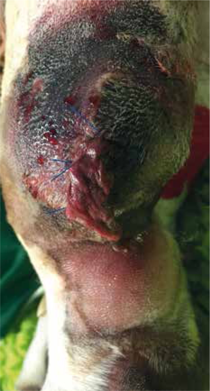
From the moment that a patient arrives at the practice, the risk of exposure to potentially harmful flora is prominent. The two types of flora that can become problematic are resident and transient flora. Resident flora lives on normal skin and provides protection from other pathogens, whereas transient flora is acquired bacteria and does not normally colonise skin (Gregory, 2005). Exposure of a surgical wound to either resident or transient flora can result in colonisation of the wound and development of clinical signs of infection. In addition to exposure of the open wound to bacteria, the risk of SSIs is significantly elevated with increased surgical time, increased persons present during surgery, and a dirty surgical wound (Eugster et al, 2004). Therefore, the efficacy of the surgical team in preparing the patient, and the standard of practice protocols regarding wound management, can directly impact the risk of SSI contraction. Figure 1 demonstrates SSI contraction post-caudectomy in an adult canine patient; note dehiscence of the wound and presence of exudate.

Wound classification: clean versus dirty wounds
Wounds are generally categorised into four classifications (see Table 1) Clean wounds provide minimal risk of infection, whereas dirty wounds may already display signs of infection or tissue damage and are significantly contaminated, for example, a degloving injury resulting from trauma such as a road traffic accident. Foreign matter and bacteria are likely to have adhered to the wound, eliciting an inflammatory response, and the risk of SSI following surgery is increased. It is possible that dirty wounds will be managed as open wounds initially due to tissue damage or loss, and the increased risk of SSI development once closed. These wounds may need daily irrigation and debridement, as well as aseptic handling until surgical closure is a viable option. Therefore, wounds must be handled appropriately depending on their classification and in such a way that risk of SSI is reduced to a minimum.
| Clean | Typically clean wounds are those created under aseptic conditions, for example, a surgical incision. Incisions into contaminated tracts are not included |
| Clean contaminated | Similarly to clean wounds, these wounds are made under aseptic conditions, however, they enter a contaminated tract, such as the oesophagus. Alternatively, clean contaminated wounds could arise from a minor break in asepsis |
| Contaminated | Open traumatic wounds, incisions into contaminated or inflamed areas, or wounds created under aseptic conditions where a severe break in asepsis has arisen, for example cystotomy resulting in some urine contamination of the abdominal cavity |
| Dirty | Wounds where clinical signs of infection are present, or traumatic wounds typically >12 hours post-incident where bacterial colonisation is assumed. For example, bite wounds |
Personal hygiene
Peri-operatively, clean and dirty wounds must be handled with diligence to reduce the transfer of bacteria. Registered veterinary nurses (RVNs) are aware of the importance of infection control and should maintain high standards of personal hygiene. Although some evidence shows little difference between the bacterial load of bare, and non-bare skin post-hand washing, a strict bare-below-the-elbows policy can reduce the surface area for bacterial colonisation (Mann, 2016). At the time of surgery, individuals must adopt stringent aseptic techniques when preparing the surgical site to minimise the risk of contamination of the surgical field. Effective hand hygiene should be demonstrated using the World Health Organisation (WHO) handwashing technique (Mann, 2017), although practices often experience barriers to this, such as the busyness of staff (Mann 2018). Compliance of staff is imperative, and it must be remembered that all registered veterinary professionals take an oath at the time of graduation to do their best for their patients, and to maintain and improve welfare standards; therefore, protocols such as hand hygiene, are not optional. Effective hand hygiene can be achieved using either antiseptic soap or hand gel, such as Sterillium® (Hartmann). WHO have found that the use of hand gels with high alcohol content (60–80%) are in fact more effective than traditional soap and water solutions (WHO, 2009), which is important to consider when handling surgical patients. Once hands have been effectively cleansed, personal protective equipment, such as disposable gloves, should be worn to prepare the surgical site. It must be expressed that hand hygiene is as important pre-surgery as post-surgery. By following these procedures, the risk of transfer of pathogens to the wound and the subsequent contraction of an SSI, is reduced.
Surgical safety checklists (SSCs)
Not only does the implementation of SSCs comply with clinical governance to improve standards of patient care, it can increase efficacy across theatre practice. Implementation of the use of these checklists has been widely successful in human medicine, with one study recording a reduction in SSIs by 50% (Haynes et al, 2009), and the WHO Safe Surgical Checklist contributed to a fall in mortality of post-surgical patients from 1.5 to 0.8% (Oxtoby and Mossop, 2016). Furthermore, SSCs increase efficiency of the theatre by improving communication and teamwork, and provide an organised approach to theatre running (Young, 2018). This combination leads to reduced surgical time, and thus can be a vital implementation to the practice in reducing the risk of SSIs. In human medicine, one study in the United States evaluating the implementation of a checklist for placement of central intravenous lines achieved reduction of SSIs from 11.3/1000 to 0/1000 patients (Oxtoby and Mossop, 2016), showing the positive potential for implementation. In addition to this, SSC implementation has been successful in reducing the occurrence of ‘never events’; events that should never occur, such as failure to administrate pre-operative antibiotics (Clapham, 2015). This is an example of an imperative step before many surgeries, such as those with a high risk of sepsis. Ensuring all peri-operative treatments such as these have been given, is yet again a step towards reducing the risk of SSIs.
Although there may be significant differences between veterinary practices, implementation of SSCs can be beneficial for all by providing a standardised approach to each surgery.
Essential checks for any SSC should include:
Some larger referral hospitals may have additional criteria, such as more specific equipment checks, use of suction and any blood products available and used. Tailoring a SSC to reflect individual practice variation can ensure relevance and can thus increase compliance among staff.
Post-surgical wound management
A significant part of the surgical ward RVN's role is to monitor postoperative patients for signs of local or systemic infection. Local indicators of SSIs include oedema, erythema, exudation and pain. Figure 2 demonstrates these indicators in addition to soft tissue escape. In this case, the patient had self-traumatised, likely as a result of associated pain. The most recognised indicators for systemic infection include pyrexia, as well as lethargy and depression. In the hospital environment, patients should be observed every 4–6 hours to allow for deterioration of demeanour and the wound site to be detected (Breton, 2012). Vital signs such as heart rate and temperature should also be taken regularly. However, it must be recognised that clinical signs of infection can develop as long as 14 days postoperatively (Elishar, 2008). By this point, patients have often been discharged, and therefore postoperative management must be achievable at home, by clients.
For critical patients hospitalised for >24 hours, a temperature should be taken at least once per day to allow for spikes to be identified in the days following surgery (Humm and Kellett-Gregory, 2016). In addition, changes in heart rate, respiration rate, blood pressure and demeanour should be noted and communicated to the team. Local inflammation linked with SSIs can cause pain, often indicated by patient interference, vocalising, lameness or guarding of the affected area. Consequently, recognised pain scoring systems such as the Glasgow Composite Pain Scale (canine and feline modifications), Feline Acute Pain Scale and the Rabbit Grimace Scale should be utilised to enable identification of early changes consistent with SSI development.
For patients hospitalised for <24 hours, regular checks should also be carried out. However, it is unlikely that clinical signs of SSI will present prior to discharge. A recent study identified SSIs in 2.83% of patients following discharge; no SSIs were detected prior to discharge and the mortality of these patients was 8.3% after development of sepsis (Garcia Stickney and Thieman Mankin, 2016). Therefore, this not only highlights that client education is of utmost importance, but that follow-up procedures, such as telephone calls or face-to-face appointments, are essential in the early detection of SSIs. With regard to prevention, owners must be aware of the importance of averting patient interference and effective wound monitoring. Information sheets detailing this are invaluable due to the effect of information overload during the discharge appointment. Clients are often put off by buster collars, therefore stocking alternatives, such as body suits, may enable increased compliance and thus reduced occurrence of SSIs.
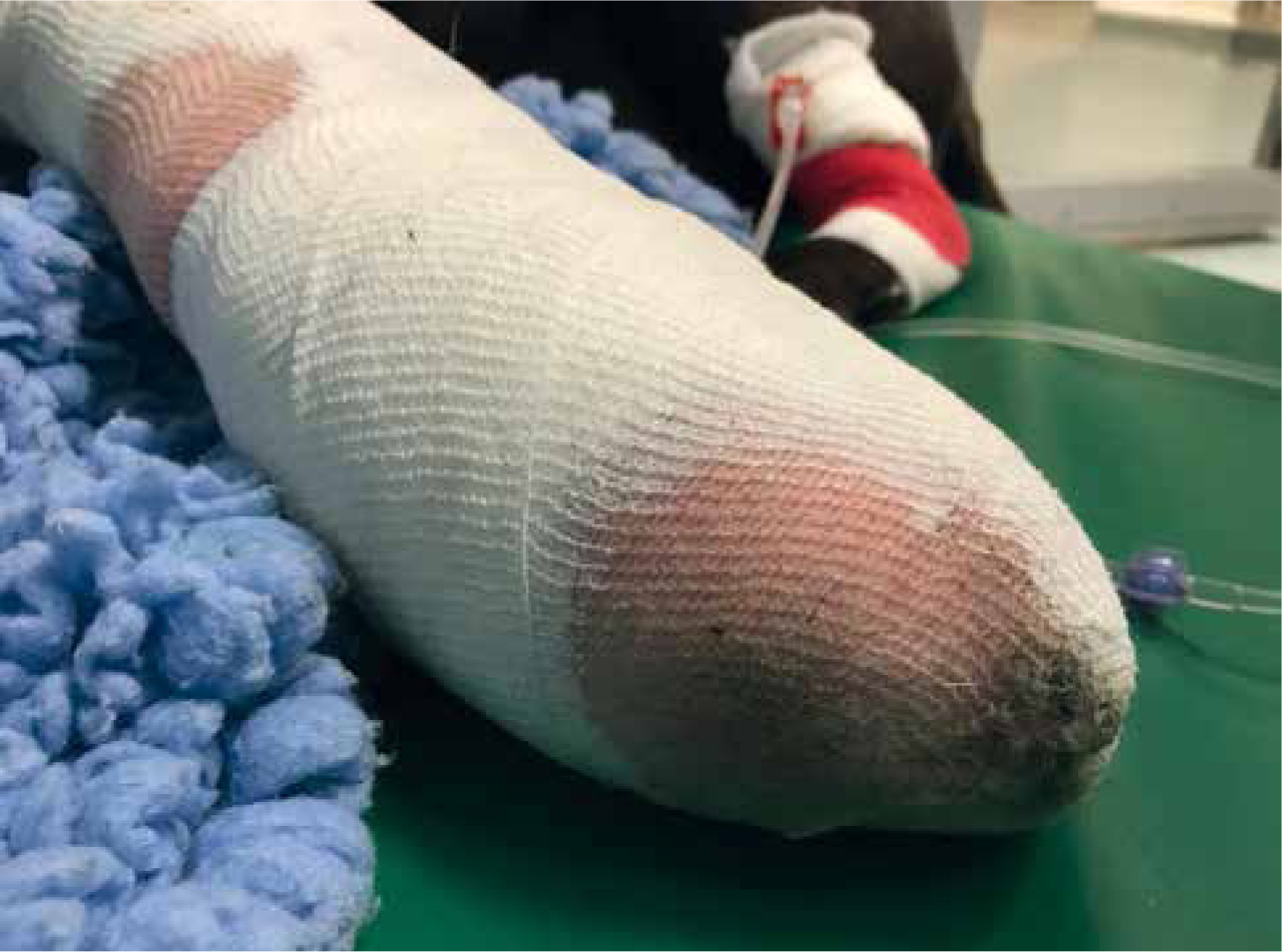
The use of dressings may be indicated to prevent transfer of transient flora from the environment to the wound, and in the case of infection, from the affected wound to other patients during hospitalisation. For clean wounds, simple dressings, such as passive dressings, are ideal as these minimise interaction with the wound and for their absorptive properties. All dressings, including bandages, must be monitored for strikethrough: changes in colour, odour and volume can all indicate SSIs (Figure 3).
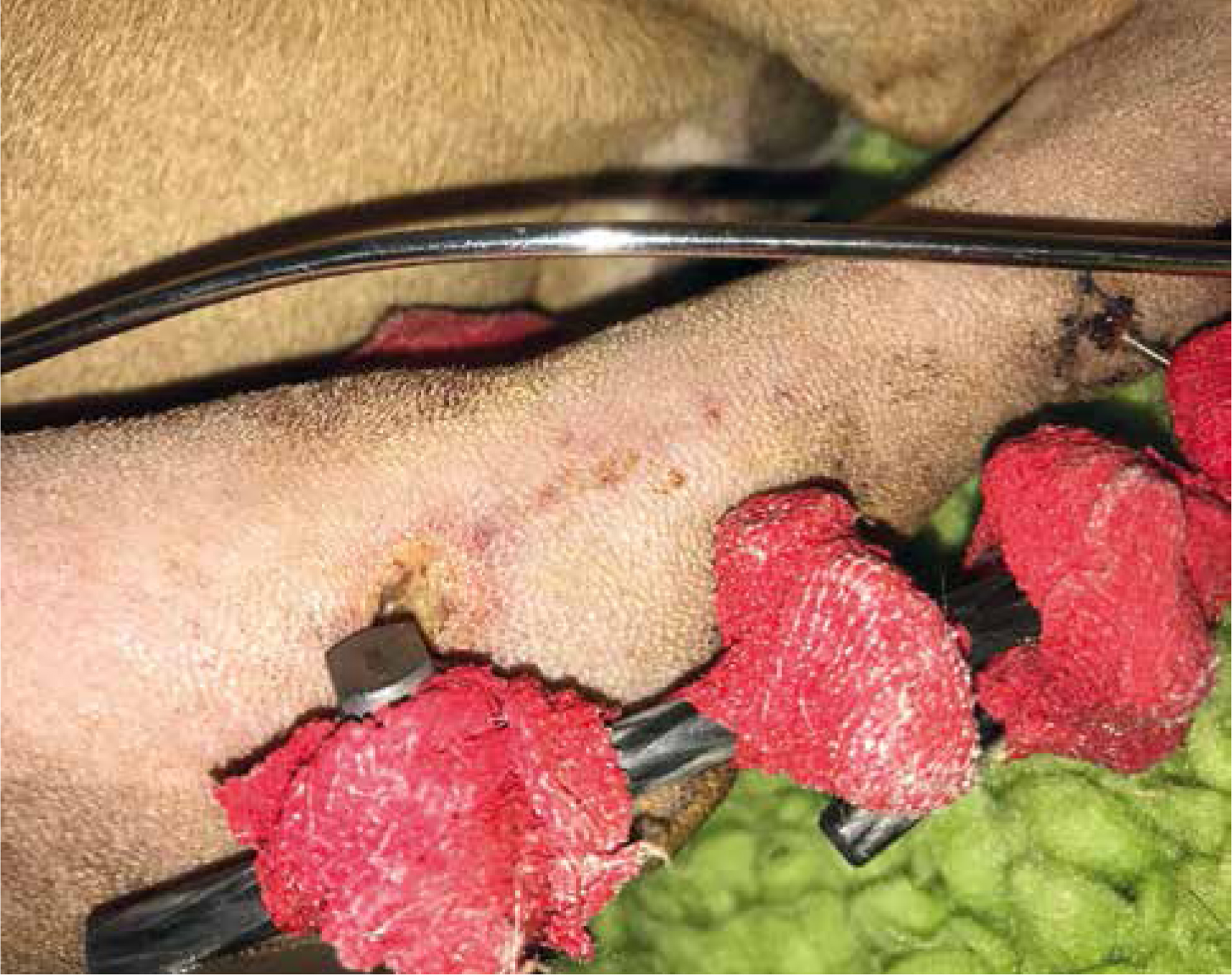
Extra care must be taken when handling drains and orthopaedic structures such as external fixators. These provide tracts for bacteria to ascend and to enter the body and result in infection. Therefore, strict asepsis must be demonstrated when handling. For drains, monitoring for changes in exudative characteristics, such as odour and colour, in addition to recognising increased production is crucial. For external fixators, monitoring the entry and exit sites of pins for signs of pin-tract infections is critical. Figure 4 shows a pin-tract infection following fracture repair of the femur. Note the yellow-green exudate pooling at the most proximal pin.
Reducing the risk of ophthalmic SSIs
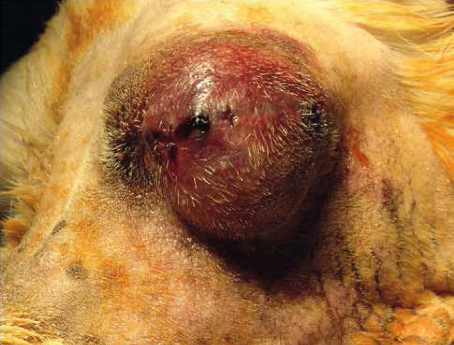
Veterinarians have been seeing ophthalmic patients for many years, however, recently, development of treatment protocols in this field has accelerated. Ophthalmic patients at risk of SSIs include those postoperative from phacoemulsification, ulcer repair and foreign body removal, to name a few. Clinical signs include inflammation, discharge of varying appearance, pain and dehiscence. Figure 5 shows oedema and erythema post-enucleation in a canine patient.
If infection is suspected prior to surgery, a diagnostic cytology slide should be prepared to allow identification of bacteria for accurate antibiosis to be achieved. Preparation of the surgical site should include a wash of a 50% iodine solution, as opposed to scrubbing, which would cause ocular trauma, and chlorhexidine which is a corneal irritant (Roberts, 2013). Managing the surgical wounds of these patients can be challenging due the fragility of the eye and the potential for rapid deterioration, especially where grafts have been placed. It is therefore the responsibility of the RVNs involved to monitor the site regularly, administer medications and flush with antiseptic solutions where indicated, while maintaining strict asepsis.
Antibiotics
Antibiotic prophylaxis in the veterinary world is contributing to the frighteningly real problem of antibiotic resistance, and a sharp rise in the occurrence of multi-drug resistant infections (MDRIs), such as methicillin-resistant Staphylococcus aureus (MRSA) and Staphylococcus pseudintermedius (MRSP) (Roberts, 2013). Contraction of these MDRIs can be devastating for the veterinary team, patients and clients. Therefore, it is important for RVNs to recognise the appropriate use of antibiotics and as valued members of the veterinary team, discussions regarding their use are those that RVNs can, and should, be involved in.
Conclusion
In conclusion, SSIs can be devastating. Therefore, RVNs play an important role in prevention and monitoring for signs of infection. Implementation of exceptional hygiene protocols, the use SSCs and a solid understanding of how to handle different wounds is essential for the prevention of SSIs.