
Rehabilitation techniques can play an important role in management of the canine hip dysplasia patient to complement more conventional medical and surgical options. Discussion of the techniques described in this article will provide owners with a full, well-rounded understanding of the benefits rehabilitation can provide.
It is important to ensure adequate pain control is in place before initiating any exercise programme to prevent aggravation of clinical signs.
Rehabilitation goals
The goals of rehabilitating a patient with hip dysplasia include:
A number of techniques can be employed in practice by the veterinary nurse, at home by the pet owner, or during referral appointments by the rehabilitation therapist. A number of these techniques will be described below.
Thermotherapy
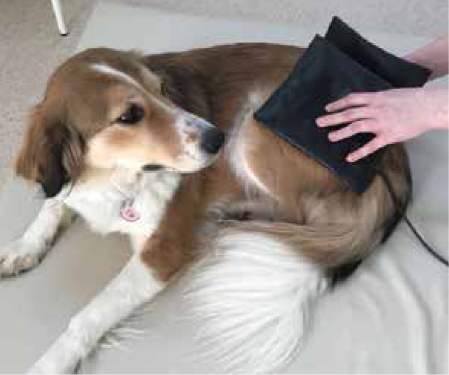
Both cryotherapy (easily administered through application of a cold pack wrapped in a damp cloth or towel to prevent insulation to an acutely painful joint for 5–15 minutes) and application of low-level heat can play a part in pain management in the patient with hip dysplasia. Cryotherapy can be used for up to 3 days following an acute flare up, applied as described above, every 4–6 hours.
Cryotherapy causes vasoconstriction which assists in the control of tissue oedema, and slows enzyme activity, which in turn decreases the activity of the inflammatory pathways, controlling pain, swelling and inflammation (Millis and Levine, 2014).
A microwavable heat pack can easily be used at home or in practice, and is best applied to the soft tissues rather than directly over a painful joint for optimum results, as it has been demonstrated that heat therapy provides pain relief, reduces muscle spasm and increases soft tissue elasticity (Carver, 2016), and so is an excellent precursor to soft tissue techniques, such as massage. Heat packs should be applied for around 15–20 minutes and can be used daily.
Soft tissue massage
Application of soft tissue massage may decrease myofascial pain and muscle tension (Dycus et al, 2017). This can be applied to the hip dysplasia patient on a daily basis for anything from 5–20 minutes, depending on patient compliance, as a combination of long and short massage strokes, directed along the spine, over the gluteal, quadricep and hamstring muscles. These massage techniques also provide sensory stimulation and so enhance proprioception, and relax the tissues and patient via endorphin release (Millis and Levine, 2014). Effleurage increases circulation and so can be used to enhance blood flow to a desired area to enable efficient oxygen delivery to the tissues and assist in the removal of waste products (Griffiths, 2014).
Passive range of motion exercises (PROM)
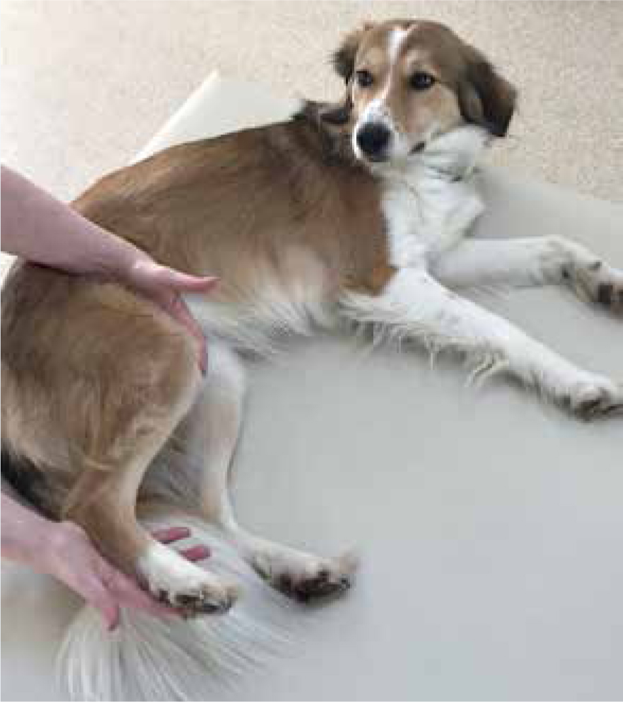
PROM exercises involve moving the joints passively by the therapist slowly through their available range, until natural resistance is met. In practice, this is often best achieved with the patient relaxed and lying in lateral recumbency, with the upper limb supported by the therapist, taking care to keep the limb in a level (sagittal) plane, as per Figure 1. Starting distally with the digits and moving up through the tarsus, stifle and then hip; each joint should be moved through around ten flexions and extensions, in three sets, twice daily for maximum results. As maintaining joint range of motion is much more effective than trying to increase it, this should be started as early in the disease process as possible. Other benefits of PROM exercise include enhancing proprioception and increasing circulation (Carver, 2016).
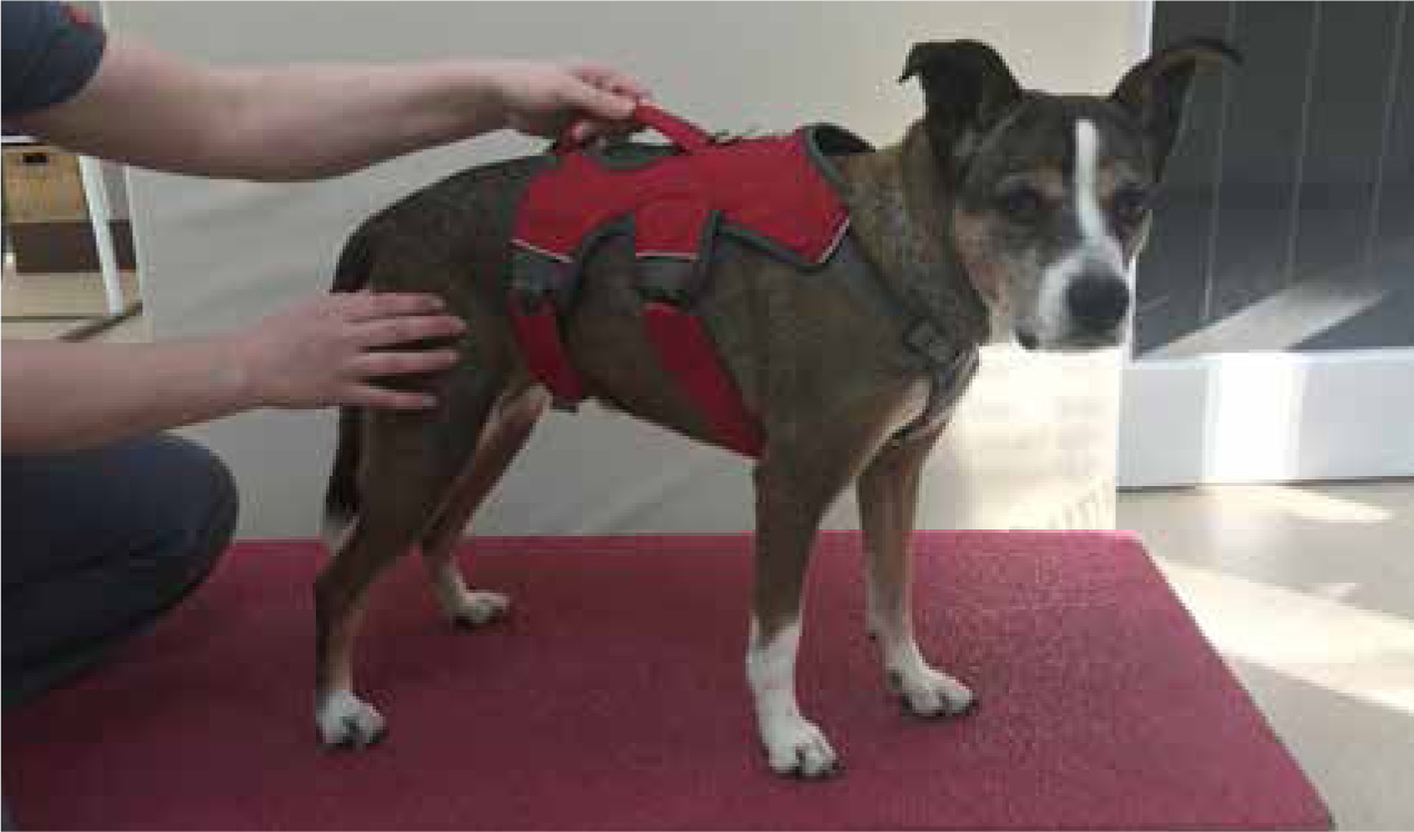
Active range of motion exercises (AROM) and strengthening exercises
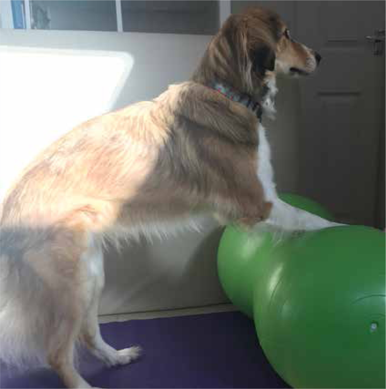
AROM differs from PROM in that the patient is responsible for ‘actively’ moving the joints through their natural range, rather than being performed by the therapist passively. As well as the benefits of PROM, most forms of AROM initiate muscle contraction and so double up as strengthening exercises (Prydie and Hewitt, 2015). Examples include:
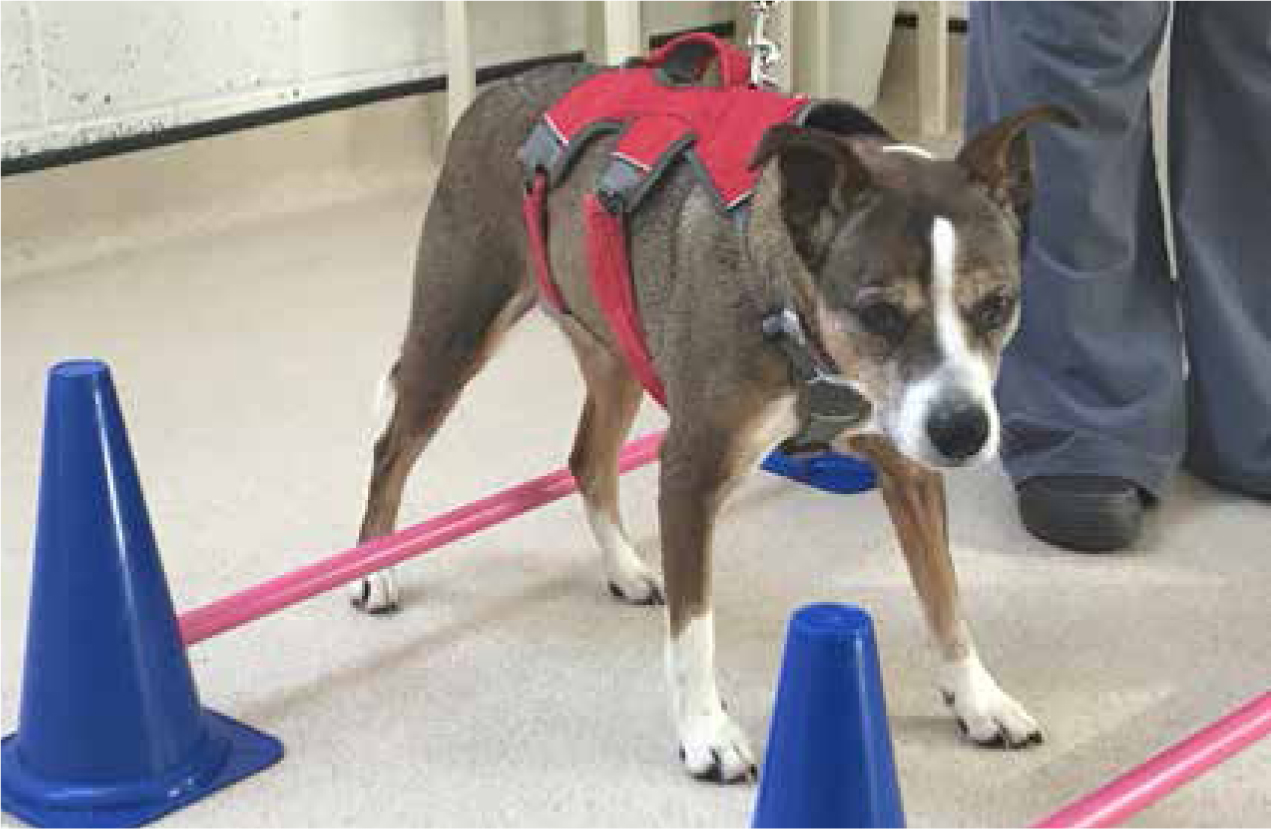
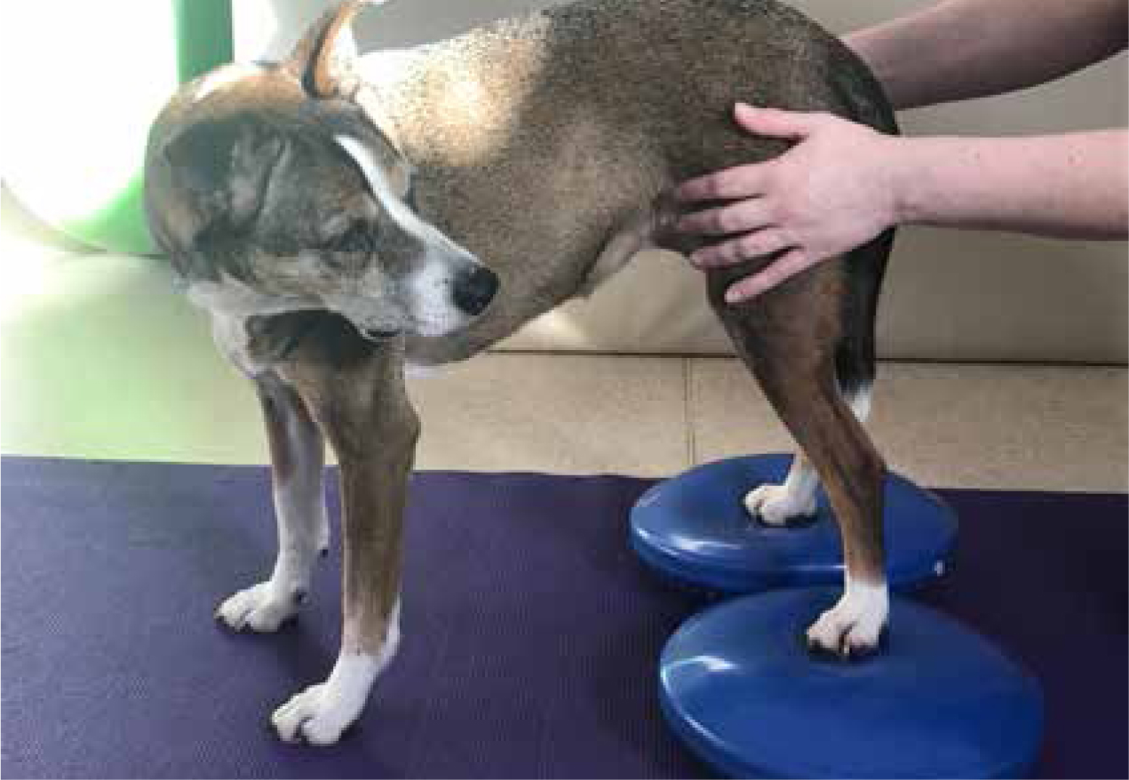
Proprioceptive enhancing techniques
Proprioceptive deficits are common in humans suffering from joint disease, and can lead to balance and coordination issues. Techniques to enhance proprioception should be incorporated as part of a rehabilitation programme for hip dysplasia patients in order to prevent or address sensory deficits (Dycus et al, 2017).
Aquatic therapy
Hydrotherapy techniques involve exercising in a water environment to provide adequate buoyancy to support the patient's bodyweight, and therefore reduce impact on the joints (Prankel, 2008), while building muscle. Exercising in water also improves the range of motion of joints (Sharp, 2008).
Referral of the patient to a hydrotherapy centre, particularly with access to an underwater treadmill, would be ideal. This would be utilised with the water level at the height of the hip joints to provide support (Wong, 2011), but without being so deep as to encourage swimming rather than walking, which is a functional gait pattern and encourages hip extension (McGowan et al, 2007). Sessions are normally initiated once or twice weekly. A useful outcome marker is to measure the girth of each hindlimb before the first session, and after a block of, for example, 10 sessions; effective use of the muscle will increase the girth of the hindlimb. This technique can also be applied to any exercise programme to measure patient progress, but as with goniometry, it is most accurate when performed by the same person each time (Rothstein et al, 1983).
Electrotherapy
Pulsed electromagnetic energy (PEME) alters the transfer of pain signals, via the hyperpolarisation of the cell membrane, which prevents the depolarisation required to produce a nerve impulse and so the delivery of a pain signal. This is achieved through altering calcium ion transport through the cell membrane by creating a magnetic field by directing an electrical current through a coil of wire. The result is an analgesic effect which has been demonstrated to assist in the management of pain, in both the acute (Stocchero, 2015) and chronic (Ryang, 2013) form. Pads applied directly over the hip joints (Figure 6) are often used by the rehabilitation therapist either before or after therapeutic exercise techniques.
LASER therapy may also be applied to provide analgesia and anti-inflammatory effects (Griffiths, 2014). This is achieved via the delivery of photons to the cells, which trigger a number of reactions within the mitochon dria. Enzymatic production of adenosine triphosphate (ATP), the energy unit of the cell, is stimulated, increasing the speed of cell metabolism and function, including DNA production and healing and repair of the cell. ATP can also function as a neurotransmitter, altering pain modulation (McGowan et al, 2007).
Environmental adaptations in the home
Advising the pet owner on adapting the home environment to improve patient quality of life can have a dramatic impact (Tanner, 2018). Non-slip mats used on tiled or highly polished floors can prevent slipping or splaying accidents and improve patient confidence when moving around the home. Using aids such as ramps or pet steps in and out of the car, and onto furniture if the pet is used to access, will reduce the landing forces and so impact from jumping up and down, and prevent frustration or accidents from failed attempts at jumping. Limiting access to stairs by use of baby or dog gates may be useful in patients with pain or hindlimb weakness in the early stages of rehabilitation, again to prevent accidents from attempts particularly when the owner is not at home. Provision of a deep bed with low-front easy access will provide plenty of support for the joints without the patient having to climb over the sides.
Conclusion
The veterinary nurse can play a vital role in the management of the hip dysplasia patient, from recognising early signs of pain, to assisting with weight management, initiating and discussing exercise programmes and environmental adaptations for the owner at home, and performing gentle physiotherapy techniques in practice. All of these techniques allow a global, multimodal approach to management of a complex, extremely common orthopaedic disease, which can have a huge impact on patient welfare and quality of life.