

Canine heart disease is commonly seen in veterinary practice. Presentations that veterinary nurses might recognise range from the healthy dog that appears for annual vaccination with a heart murmur, to a patient that is admitted with a concurrent problem, such as cruciate surgery, or the patient that presents with congestive heart failure (CHF), which can be either acute or chronic.
Heart disease in dogs can have different origins, whether it is present from birth (congenital), develops at a later age (acquired) or is the result of an arrhythmia (arrhythmogenic). This article will focus on the most common forms of acquired heart disease seen in veterinary practice:
- Mitral valve disease (MVD)
- Dilated cardiomyopathy (DCM).
MVD has been shown to account for up to 70% of canine cardiac disease in veterinary practice (Det-weiler and Patterson, 1965; Häggström et al, 2004; Borgarelli and Häggström, 2010). MVD is characterised as a progressive disease, with a classic leftsided apical heart murmur, and is often seen in smaller breeds of dog. DCM is a myocardial disease characterised by ventricular dilatation, seen in medium- to large-breed dogs. Over time, the ventricle walls become unable to relax and contract properly, thereby reducing cardiac output. A diagnosis of DCM can be more challenging because it can have a primary myocardial origin, or can be the result of secondary diseases, such as nutritional or hormonal deficiencies.
The cardiovascular system
Normal cardiovascular function provides the body with nutrients and removes metabolic waste. The heart is the pumping force behind this system, and is divided into two sides to prevent mixing of oxygenated and deoxygenated blood. Deoxygenated blood returns to the right atrium via the vena cavae, across the tricuspid valve and leaves the right ventricle through the pulmonary artery. The pulmonary veins return oxygenated blood to the left atrium, which crosses the mitral valve and enters the systemic circulation through the aorta.
Approximately 75% of blood volume is found in the systemic circulation (Ware, 2007). The pressure required to push blood around the systemic circulation is therefore much higher. This is known as systolic pressure. Pulmonary circulation, or diastolic pressure, is about one seventh of the total mean blood pressure (Ware, 2007). The difference in these pressures is reflected in intracardiac pressures, where the left-side pressures are much higher than the right. The pumping action of the heart is influenced by:
- Electrical activation
- Ability of the ventricles to pump (relax and contract)
- Degree of filling during diastole and level of atrial contraction
- Heart rate.
Heart disease can interfere with these factors and the heart may therefore be unable to meet the cardiac output demands needed to maintain blood pressure (Box 1). If blood pressure becomes compromised, the body tries to compensate by activating several mechanisms in an attempt to maintain adequate blood flow and subsequent tissue oxygenation. However, these mechanisms can accelerate the process of heart failure. For example, when cardiac output decreases, the renin-angiotensin-aldosterone system (RAAS) and adrenergic system increase heart rate and retain fluid to increase plasma volume.
Box 1.Cardiovascular regulation formulaeThese two formulae explain how the circulatory system is tightly regulated. If blood pressure falls, compensatory mechanisms are activated to normalise blood pressure. This can be life saving, for example in cases of acute haemorrhage, but can have deleterious cardiovascular effects if sustained.
- Blood pressure = Systemic vascular resistance (the resistance that the left ventricle must overcome to pump blood around the systemic circulation).+ cardiac output
- Cardiac output = Stroke volume (the amount of blood pumped by one ventricle in each contraction).+ heart rate
Cardiac causes of compensatory activation include:
- Systolic function: impairment of the heart-pumping mechanism to eject blood into the arteries during systole; occurs in DCM, when the ventricular myocardium is weak and cannot pump effectively
- Volume overload: an increase in the volume of blood that must be pumped by the heart; most commonly occurs with MVD, in which blood flows back into the left atrium as a result of the leakage of the mitral valve
- Pressure overload: an increase in pressure on the heart when the ventricles pump blood into the arteries. This can be owing to a narrowing (stenosis) of the aortic or pulmonic valves or to systemic or pulmonary hypertension
- Diastolic dysfunction: impairment of the filling of the ventricles during diastole; commonly occurs in mitral valve stenosis and pericardial effusion
- Arrhythmias: can impair cardiac filling and output owing to an increased or decreased heart rate.
Cardiac disease in dogs can take a number of different forms. The most common are the acquired diseases, MVD and DCM. Congenital diseases can be seen, and are a result of either malformation of one or more areas of the heart or great vessels, persistence of normal fetal structure following birth or valvular abnormalities. The most prevalent congenital diseases in the UK are patent ductus arteriosus, aortic stenosis and pulmonic stenosis (Martin and Dukes-McEwan, 2010). Dogs can be affected by many arrhythmogenic diseases, which may or may not have a cardiac cause.
Box 2.Other names for mitral valve disease
- Mitral valve disease
- Degenerative mitral valve disease
- Myxomatous mitral valve disease
- Chronic mitral valve fibrosis
- Chronic degenerative valvular disease*
- Chronic valvular disease
- Endocardiosis*
*Names including both the mitral and tricuspid valves
Mitral valve disease (MVD)
There are a number of names used for valvular acquired cardiac disease in dogs, which can be confusing. A list of commonly used names is provided in Box 2, although this is not an exhaustive list. When considering terminology, it is important to recognise that some of them refer to just the mitral valve, while others relate to both mitral and tricuspid valves. Degeneration of the left atrioventricular valve is the most commonly diagnosed, but degenerative changes can affect all heart valves. Buchanan (1977) found the following degenerative changes affecting heart valves:
- Mitral valve alone: 62%
- Mitral and tricuspid valves: 32.5%
- Tricuspid valve alone: 1.3%.
Although this is a historical study, these figures are still cited and used by leading cardiologists (Fox, 2012). For the purpose of this article, the term MVD will be used throughout.
MVD is characterised by degeneration and fibrosis of the valve apparatus. This thickening means that the valve leaflets cannot close as they should. Therefore, when systole occurs and blood is forced through the aorta, some blood will pass back through the mitral valve and back into the left atrium. This is known as a regurgitant jet, and this mixing of blood is when a heart murmur can be detected. Mitral valve regurgitation can affect cardiac output (owing to less blood passing through the aorta), and when this happens, compensatory mechanisms are activated to increase ventricular stroke volume.
Long-term activation of these compensatory mechanisms can lead to atrial and ventricular dilatation because of increased plasma volume. This, in turn, exasperates leaking across the valve, as the mitral valve cannot stretch to accommodate the dilatation that has occurred. Furthermore, as MVD is a progressive disease, the mitral leaflets can naturally degenerate further, causing increased stress and structural damage. This cycle can continue over years as the leaflets become more and more distorted. Eventually, it can lead to rupture of the chordae tendineae, further increasing the volume of regurgitant blood. When advanced or severe, MVD can result in decreased cardiac output and increased pressure in the atria, as well as a build-up of pressure in the venous system. Recent studies have been useful in showing prognosis statistics of dogs with MVD. Borgarelli et al (2012) showed that in a study of 256 dogs, 60% lived for more than 5.5 years after diagnosis. Prior to this, Borgarelli and Häggström (2010) reported that 30% of dogs with mitral regurgitation progressed to heart failure and Borgarelli et al (2008) showed that 50% of dogs with severe heart failure died within 9 months as a result of MVD. What these studies indicate is that prognosis for a dog with MVD is considerably worse if they progress to heart failure.
Epidemiology
MVD can be seen in all breeds, but it is most commonly seen in small- to medium-sized breeds. The Cavalier King Charles Spaniel (CKCS) is particularly susceptible to the disease, with one report showing 90% of CKCS diagnosed with MVD by the age of 10 (Borgarelli and Häggström, 2010). Other breeds also predisposed are Poodles, Whippets, Terriers and Chihuahuas. Additionally, studies have shown that male dogs are affected at an earlier age, and tend to present with a more aggressive form of the disease (Häggström et al, 2004; Egenvall et al, 2006).
Aetiology of MVD
The cause of MVD is not yet fully understood. It is believed that in affected dogs, the connective tissue within the valve is defective, which results in the valve degenerating over time (Häggström, 2010). There have been studies investigating molecular pathways, (Oyama and Chittur, 2006) and levels of serum serotonin (Arndt et al, 2009), which have shown some interesting trends. Madsen et al (2011) found a genetic link between two loci and MVD. Parker and Kilroy- Glynn (2012) have linked small body size with an increased likelihood of MVD. Only fairly recently was MVD shown to be an inherited disease in CKCS and Dachshunds (Häggström, 2010). This does not, however, explain how or why other small-breed dogs are affected.
Dilated cardiomyopathy (DCM)
DCM is the most common acquired myocardial disease in dogs (Sisson et al, 1999). Cardiomyopathy is a term used to describe an inadequately functioning heart muscle, and is characterised by heart-chamber dilatation. When a heart is affected with DCM, the ventricle fails to expel all the blood in systole and, therefore, as cardiac output declines, compensatory mechanisms are activated. As with MVD, this extra plasma volume leads to progressive dilation of the ventricle. Chronic activation of the RAAS and adrenergic system can contribute to progressive myocardial damage, which will eventually lead to CHF. Tachyarrhythmias are commonly associated with DCM (Ware, 2007). It is common for both right- and leftsided heart failure to occur with DCM (Tidholm et al, 2001). If the heart is unable to maintain cardiac output, and is not compensated sufficiently, forward or low-output failure can also occur. Poor forward blood flow can lead to poor coronary perfusion, further progressing myocardial dysfunction and provoking arrhythmias, due to the inadequate blood flow. Prognosis for dogs in CHF as a result of DCM is guarded to poor. Ware (2007) references Monnet et al (1995) and Calvert et al (1997a), who placed the probability of survival for 2 years between 7.5–28%. Martin et al (2009) reported that 50% of dogs with CHF resulting from DCM died within 4 months. Since then, however, the PROTECT study, reported in 2012, showed a benefit for pimobendan to delay the onset of CHF or death in Dobermans with preclinical DCM (Summerfield et al, 2012).
Epidemiology
Idiopathic DCM is most commonly diagnosed in large- and giant-breed dogs, although Spaniel breeds (particularly the English and American Cocker Spaniel) can also be affected. A retrospective study conducted at Purdue University from 1986–91 showed that Deerhounds, Dobermans, Irish Wolfhounds, Great Danes, Boxers, and Newfoundlands were overrepresented. There does not seem to be a gender predisposition to DCM, but males are more likely to present in CHF and at an earlier age than females (Dukes-McEwan, 2010). A genetic link has been found in Irish Wolfhounds (Vollmar, 2000) and in Dobermans (O'Grady and O'Sullivan, 2004), and an autosomal dominant inheritance pattern has been reported in Boxers with an associated ventricular arrhythmia (Meurs et al, 1999). DCM is more often diagnosed in middle-aged dogs, but a wide age range has been reported, some as young as a few weeks old (Dukes-McEwan, 2010).
Aetiology of DCM
The most common form of DCM, primary cardiomyopathy, is referred to as ‘idiopathic’ because the cause of the myocardial disease is unknown. There are other causes of cardiomyopathy in dogs, known as secondary cardiomyopathies. Hypothyroidism, taurine deficiency and the chemotherapy drug, doxorubicin, can all lead to DCM. There are two phases of idiopathic DCM described. During the first phase, preclinical or ‘occult’, DCM is often clinically silent. During the second phase, ‘overt’, DCM can occur suddenly, and will commonly present with signs such as lethargy, tachypnoea, cough and anorexia (Ware, 2007). Syncope and sudden death can occur in association with ventricular arrhythmias. It has been documented that Dobermans may have ventricular arrhythmias long before clinical signs are noted (Calvert et al, 2000).
Histopathology of DCM
Histopathology has found that DCM has two distinct types. In most dog breeds, and particularly in giant dogs, post-mortem analysis has shown that myocardial cells have a wavy and narrowed appearance (Tidholm et al, 1998). Another type of post-mortem analysis is characterised by myofiber degeneration, myocyte atrophy, cell destruction, fatty infiltration and fibrosis. This pattern has been described in Dobermans (Calvert et al, 1997b) and in Boxer dogs (Dukes-McEwan et al, 2003). These are similar to the histopathological findings found in Boxer cardiomyopathy, also known as arrhythmogenic right ventricular cardiomyopathy (Dukes-McEwan et al, 2003).
Clinical signs of MVD and DCM
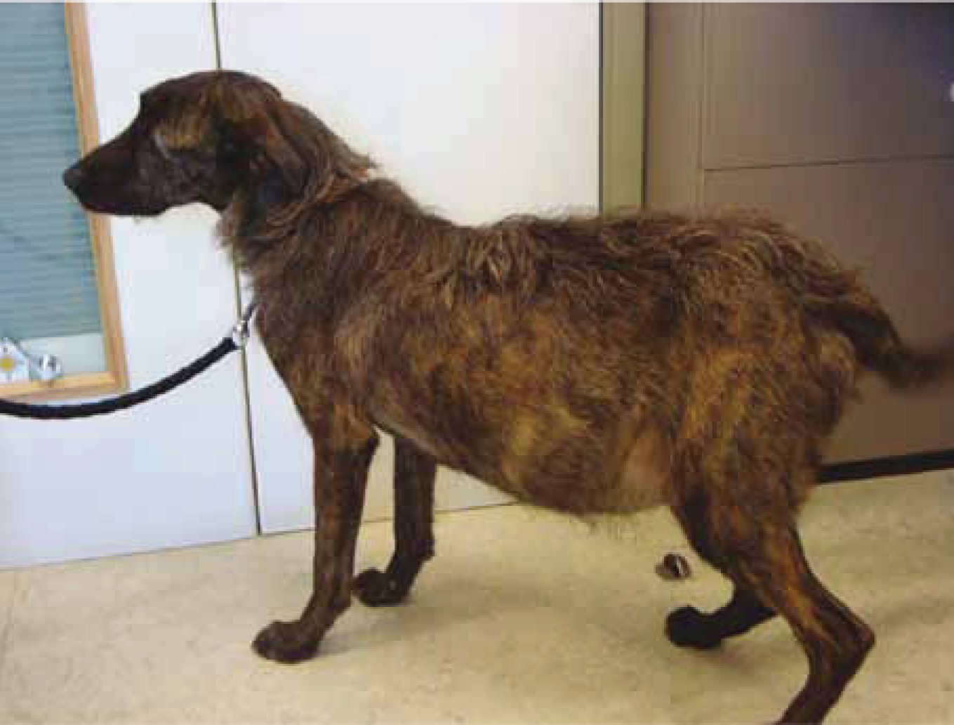
Clinical signs of MVD and DCM are similar because the left side of the heart is primarily affected (Table 1). A common finding in patients with CHF with either disease is that they become anorexic. One study showed that anorexia was a contributing factor in owners' decision to euthanise their pet with CHF in 68% of cases (Mallery et al, 1999). Cardiac cachexia may also be seen in advanced MVD or DCM (Figure 1).
Table 1. Clinical signs of MVD and DCM
| MVD | DCM |
|---|---|
| Asymptomatic | Asymptomatic |
| Left apical systolic heart murmur | Left apical systolic heart murmur |
| Signs of left-sided congestive heart failure: tachypnoea, cough, exercise intolerance, syncope, increased respiratory effort and/or respiratory distress | Signs of left-sided congestive heart failure: tachypnoea, cough, exercise intolerance, syncope and respiratory distress |
| Arrhythmias | Arrhythmias, especially in high-risk breeds such as Dobermans and Boxers |
| In severe cases, both left and right sides affected | In severe cases, both left and right sides affected |
| Poor appetite | Poor appetite |
| Some dogs (e.g. Dobermans) present with an acute onset of heart failure, with no previous signs | |
| Sudden death |
Note: clinical signs vary hugely, from asymptomatic to acute, life-threatening congestive heart failure
Diagnostic tests
The most common form of diagnostic test is physical examination, including auscultation. Not only does this allow body- and muscle-mass scoring, but also provides valuable information related to heart rate and rhythm, murmur intensity and location, and lung sounds. Thoracic radiography is essential to assess pulmonary oedema formation and to differentiate from pulmonary disease (Beardow, 2008). An alveolar lung pattern, pulmonary vein congestion and left-atrial dilation are characteristic of left-sided heart failure (Baines, 2010). Echocardiography is useful to assess individual cardiac chambers, valve function and blood flow. Electrocardiography provides information on heart rate and rhythm.
Blood-pressure measurement is an excellent way of monitoring cardiac output, as well as the effect of drug therapy. The American College of Veterinary Internal Medicine (ACVIM) (Brown et al, 2007) consensus statement on systemic hypertension stated the importance of a standardised and repeatable process for taking accurate and reliable blood-pressure readings. Blood tests are also an important diagnostic tool. Serum biochemistry can evaluate electrolyte and renal parameters, vital for patients receiving diuretics. Research has proven the efficacy of newer tests that assess cardiac biomarkers, such as cardiac N-terminal pro-brain natriuretic peptide (NTproBNP) (Oyama et al, 2008). NTproBNP is a hormone that is released in response to stretch or stress of the myocardium, and can be measured in the blood. This blood test is helpful for monitoring incremental changes in heart disease (Beardow, 2008).
Treatment of MVD and DCM
Both MVD and DCM have been surgically treated, but this has not become routine practice in veterinary medicine. Surgery is high risk and financially inhibitive for many owners. Treatment is usually not indicated before clinical signs of CHF have occurred (Fuentes, 2010). The PROTECT study, however, has recently found that treating Dobermans with pimobendan with pre-clinical DCM extended survival times (Summerfield et al, 2012). There is another study in progress, the EPIC (Evaluation of Pimobendan In dogs with Cardiomegaly) study that is assessing the use of pimobendan in pre-clinical dogs with MVD. As left-sided heart failure is the most common presentation when CHF occurs, patients are treated similarly with MVD and DCM, although they are different diseases (Table 2).
Table 2. Drug groups commonly used in the management of congestive heart failure
| Chronic heart failure | Acute heart failure | Effect |
|---|---|---|
| Diuretics such as furosemide | Diuretics | Remove excess fluid |
| Positive inotropes such as pimobendan | Positive inotropes | Improve pumping function of the ventricles |
| Neurohormonal blockade such as ACE inhibitors, spironolactone | Neurohormonal blockade | Inhibit activation of the renin-angiotensin-aldosterone system (RAAS) |
| Anti-arrhythmic drugs dependent on rhythm present | Anti-arrhythmic drugs dependent on rhythm present | Minimise impact on cardiovascular function |
| Vasodilators such as dobutamine | Counteract vasoconstriction of RAAS and adrenergic system | |
| If an underlying cause has been found (secondary DCM), appropriate treatment may be needed |
* Note: oxygen therapy may also be indicated with either disease (Atkins et al, 2009)
Key Points
- Mitral valve disease is the most common acquired cardiac disease seen in veterinary practice.
- Mitral valve disease can be diagnosed in all dog breeds, but is most commonly seen in small to medium sized dogs.
- Dilated cardiomyopathy is most commonly diagnosed in large breed dogs.
- Recent advances in veterinary medicine are suggesting it is beneficial to treat some patients before clinical signs of heart failure are present.
- Owner education is crucial.
Management and nursing aims
Nursing and management aims are closely integrated. A strong bond with owners is advised to optimise quality of life, regardless of the severity of the disease process. As breed predilections to both MVD and DCM have been demonstrated, owners of pets that are in high-risk categories should be advised to monitor their pet's weight. Owners should also be advised that when their pets are diagnosed with CHF, a regular drug regimen will be needed, and that drugs such as diuretics will lead to increased urination. Owners should also be educated to monitor respiration rate and effort, as well as monitor for any other side effects, such as vomiting, diarrhoea, inappetence, weakness or increased exercise intolerance. If these symptoms occur, owners may need veterinary advice. If CHF has been diagnosed, owners may be advised to modify exercise and diet. As discussed, progression of MVD may be a gradual process; therefore, owners should be aware of the potential longevity of the disease process. As sudden death as a result of DCM is not uncommon, owners need to be aware of the guarded prognosis of this disease.
Conclusion
Canine heart disease is common in veterinary practice as a concurrent disease, if not as the primary problem. This will only increase as patients live longer and treatments advance. Nursing and management aims should focus on building bonds with owners to improve quality of life of patients with heart disease and heart failure. Ongoing studies and improvements in veterinary medicine provide hope for susceptible breeds of both diseases.