The importance of detecting alterations in arterial blood pressure (BP) has become more widely recognized as a vital part of patient care, due to hypotension and hypertension having significant effects on patient health. Direct monitoring is proven to be more accurate than indirect methods and enables continuous measurements to be taken. This would be a distinct advantage in critically ill patients or for the detection of rapid changes in BP. As a result direct monitoring is considered to be the gold standard for BP monitoring. Most veterinary practices use indirect methods to estimate BP and make treatment decisions based on the values achieved from those methods. This highlights the importance of using a standard measurement protocol to improve accuracy and achieve repeatability of results.
Blood pressure ranges
The veterinary nurse (VN) should be knowledgeable with regards to the normal ranges of blood pressure (BP) in dogs and cats, the physiological effects of anaesthesia on BP and consequences of prolonged periods of hypotension or hypertension. During anaesthesia a mean arterial pressure (MAP) of >60 mmHg is required to ensure adequate perfusion of vital organs, if this is not maintained then signs of shock and organ dysfunction may be seen (Egger, 2007). MAP can be assessed with direct monitoring and with the oscillometric method of indirect measurement, but the Doppler method only measures systolic pressure. Systolic pressure should be >80 mmHg to prevent hypotension in anaesthetized patients (Macintire, 2010). Stepien (2010) defined hypotension in conscious patients as a systolic pressure of <100 mmHg. Hypertension results in increased workload on the heart and increases myocardial oxygen demand. Long-term hypertension can cause heart, renal disease and ocular changes such as retinal haemorrhage or retinal detachment (Love and Harvey, 2006). Hammond and Walters (1999) defined normal BP ranges as 112–192 mmHg (systolic) and 56–110 mmHg (diastolic) in conscious dogs, and 120–170 mmHg (systolic) and 70–120 mmHg (diastolic) in conscious cats.
Indirect measurement techniques
Oscillometric BP can be obtained with the use of an inflatable cuff around a limb or tail base, which is attached to a monitor. Measurement is automatic and allows detection of oscillations produced by the artery wall as the cuff deflates (Love and Harvey, 2006). Oscillometric monitors measure systolic, mean and diastolic pressure, unlike the Doppler method which only detects systolic pressure. Single measurements by this method may underestimate arterial pressure by 5–20 mmHg, meaning oscillometric BP can only be used to observe trends and accuracy may be reduced in patients under 5 kg (Love and Harvey, 2006). Oscillometric monitors have been reported to fail to produce results more often than the Doppler method (Love and Harvey, 2006).
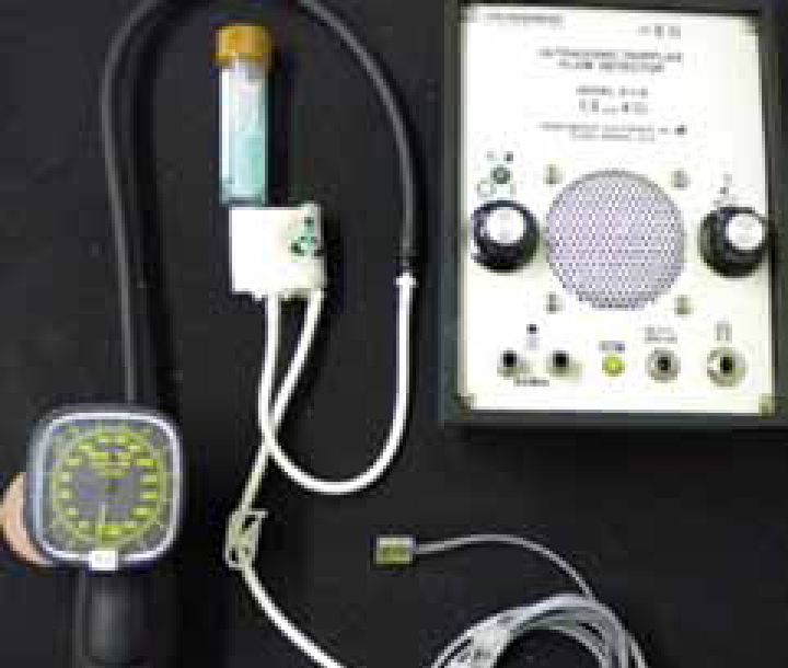
Doppler ultrasonography with the use of an ultrasound probe produces an audible sound of blood flow through an artery. Inflation until no sound is audible followed by slow deflation of the cuff until sound returns allows determination of systolic pressure (Figure 1). The Doppler method requires more skill due to manual operation and the requirement for detection of the pulse. Stepien (2010) recommends using a spirit swab or clipping the area directly over the artery if required and tolerated, followed by plenty of ultrasound gel applied to the probe to improve contact and Figure 2). Correct placement can be confirmed by the sound of blood flow, which is typically a ‘whoosh whoosh’ sound. Increased accuracy compared with oscillometric methods in smaller patients has been documented, although only measurement of systolic BP is possible (Love and Harvey, 2006).
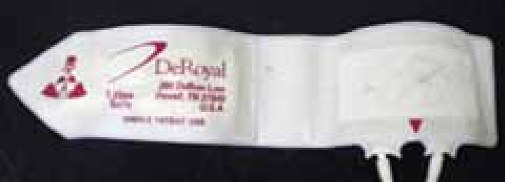
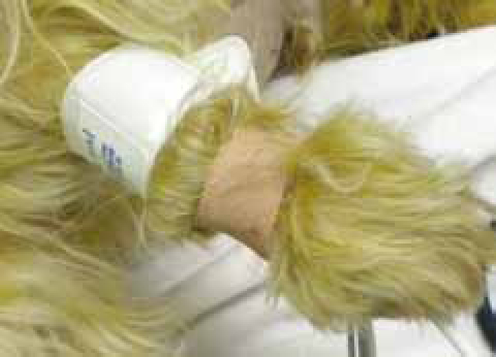
Both techniques require suitable cuff selection and correct placement (Stepien, 2000). It is recommended that the cuff width should be 30–40% of the limb circumference (Brown et al, 2007). Routine sites for cuff placement include the forelimb proximal to the carpus, the hind limb either proximal or distal to the hock or the tail base. Care should be taken to ensure the lines on the cuff are placed directly over the artery (Figure 3).
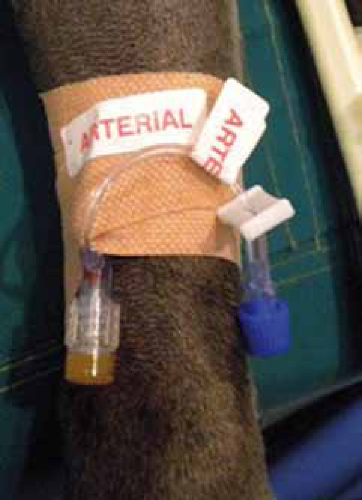
Direct monitoring
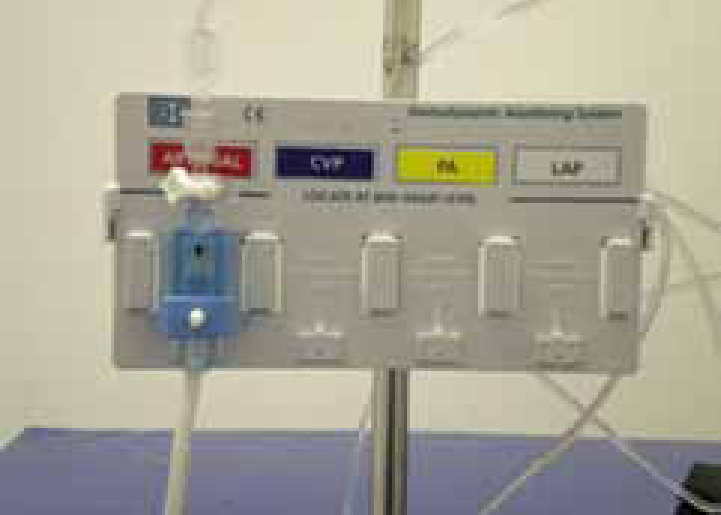
Direct monitoring requires the placement of a catheter into a peripheral artery, most commonly the dorsal metatarsal or femoral artery in smaller patients, although any accessible artery could be used (Love and Harvey, 2006). The catheter is connected to a pressure transducer with non-compliant tubing filled with heparinized saline to allow continuous measurement, which can be observed on a monitoring device (Figure 4). Most devices give continuous values and a pressure waveform can be observed. The waveform is helpful in assessing pulse quality and pulse deficits caused by an abnormal heart rhythm. Once connected to the patient the transducer must be zeroed to ambient air at the level of the right atrium (Moens and Coppens, 2007). This ensures the readings produced are accurate; regular flushing of the line will be required to ensure patency and accuracy of readings. Arterial catheters require secure taping to prevent haemorrhage due to the catheter becoming dislodged and should be clearly labelled to avoid confusion with intravenous lines (Figure 5). Most patients will not tolerate catheter placement unless critically ill or anaesthetized so this technique is unsuitable for most conscious patients.
Common technical problems
Incorrect technique is a common cause of inaccurate results, below are some problems that can be encountered.
Indirect
Patient not co-operative
Anatomical difficulties
Unable to obtain BP values/abnormal values
Unable to palpate/detect pulse
Step-by-step guide to indirect blood pressure measurement
High/low pressure not consistent with patient condition
Direct
Flat line/high pressure
Damping of trace
Abnormal readings
Importance of correct technique
A review of current literature showed correct measurement technique to be vital in ensuring accuracy of the results achieved. The American College of Veterinary Internal Medicine (ACVIM) issued a consensus statement giving guidelines for indirect measurement technique in conscious patients (Brown et al, 2007). See the step-by-step guide to indirect measurement based on the guidelines set by Brown et al (2007).
Recommendations for veterinary practice
The VN should be aware of the BP monitoring equipment available and select monitoring equipment appropriate for the individual patient. Inaccurate readings could lead to incorrect diagnosis and inappropriate management. It would therefore be appropriate to have a selection of monitoring techniques available.
Love and Harvey (2006) found that the majority of indirect monitors tend to underestimate arterial pressure but correlation with direct measurements is usually reasonable and trends can be observed. Recommendations by manufacturers should be followed to improve chances of accurate results. Consideration should be given to possible external factors that could influence the results in conscious patients such as, restraint, pain and stress. If possible these influences should be eliminated prior to measurement.
Conclusion
Nurses performing BP measurement on a regular basis should seek to improve technique and ensure compliance with guidelines set by Brown et al (2007). Repeatability of results is required and can be achieved by standardization of measurement technique. Careful interpretation and observation of trends in relation to other parameters should provide veterinary practices with a reliable diagnostic tool. Direct monitoring would still be considered the gold standard for measurement of BP and would be recommended in critical patients where accuracy is vital (Love and Harvey, 2006).